 Explore
 Validate
 Learn
Learn Western blot
 ELISA
ELISAAntibody data
- Antibody Data
- Antigen structure
- References [10]
- Comments [0]
- Validations
- Western blot [2]
- Immunocytochemistry [1]
Submit
Validation data
Reference
Comment
Report error
- Product number
- MA5-16446 - Provider product page

- Provider
- Invitrogen Antibodies
- Product name
- CD56 Monoclonal Antibody (123C3)
- Antibody type
- Monoclonal
- Antigen
- Other
- Description
- Heat-mediated antigen retrieval using a sodium citrate buffer (pH 6.0) is recommended for the staining of paraffin sections. For FACS analysis, use 10 µL of the suggested working dilution to label 1x10^6 cells in 100 µL. Mouse anti Human CD56 antibody, clone 123C3 recognizes human neural cell adhesion molecule (NCAM), otherwise known as CD56.
- Reactivity
- Human
- Host
- Mouse
- Isotype
- IgG
- Antibody clone number
- 123C3
- Vial size
- 100 µg
- Concentration
- 1 mg/mL
- Storage
- Store at 4°C short term. For long term storage, store at -20°C, avoiding freeze/thaw cycles.
Submitted references Usefulness of the octreotide test in Japanese patients for predicting the presence/absence of somatostatin receptor 2 expression in insulinomas.
Role of cd56 and e-cadherin expression in the differential diagnosis of papillary thyroid carcinoma and suspected follicular-patterned lesions of the thyroid: the prognostic importance of e-cadherin.
Variable Expression of Neural Cell Adhesion Molecule Isoforms in Renal Tissue: Possible Role in Incipient Renal Fibrosis.
CD30(+) extranodal natural killer/T-cell lymphoma mimicking phlegmonous myositis: A case report.
A Case of Distal Epithelioid Sarcoma of the Thumb Expressing Podoplanin, TLE1 and Ca 125.
Serous effusion cytology of extranodal natural killer/T-cell lymphoma.
Extraosseous (extramedullary) plasmacytomas: a clinicopathologic and immunophenotypic study of 32 Chinese cases.
Subcutaneous panniculitis-like T-cell lymphoma: a clinical and pathologic study of 14 korean patients.
Primary plasmacytoma of the cervix in a 21-year-old female patient.
Goblet cell carcinoid of the appendix: a clinicopathological and immunohistochemical study of 26 cases from southwest china.
Nakamura A, Mitsuhashi T, Takano Y, Miyoshi H, Kameda H, Nomoto H, Nagai S, Hatanaka Y, Shimizu C, Terauchi Y, Atsumi T
Endocrine journal 2016;63(2):135-42
Endocrine journal 2016;63(2):135-42
Role of cd56 and e-cadherin expression in the differential diagnosis of papillary thyroid carcinoma and suspected follicular-patterned lesions of the thyroid: the prognostic importance of e-cadherin.
Ceyran AB, Şenol S, Şimşek BÇ, Sağıroğlu J, Aydın A
International journal of clinical and experimental pathology 2015;8(4):3670-80
International journal of clinical and experimental pathology 2015;8(4):3670-80
Variable Expression of Neural Cell Adhesion Molecule Isoforms in Renal Tissue: Possible Role in Incipient Renal Fibrosis.
Marković-Lipkovski J, Životić M, Müller CA, Tampe B, Ćirović S, Vještica J, Tomanović N, Zeisberg M, Müller GA
PloS one 2015;10(9):e0137028
PloS one 2015;10(9):e0137028
CD30(+) extranodal natural killer/T-cell lymphoma mimicking phlegmonous myositis: A case report.
Yang YJ, Li YX, Liu YB, Yang M, Liu K
Oncology letters 2014 May;7(5):1419-1421
Oncology letters 2014 May;7(5):1419-1421
A Case of Distal Epithelioid Sarcoma of the Thumb Expressing Podoplanin, TLE1 and Ca 125.
Karagkounis G, Argyrakos T, Charkiolakis G, Castana O, Rontogianni D
Case reports in pathology 2013;2013:312786
Case reports in pathology 2013;2013:312786
Serous effusion cytology of extranodal natural killer/T-cell lymphoma.
Su XY, Huang J, Jiang Y, Tang Y, Li GD, Liu WP
Cytopathology : official journal of the British Society for Clinical Cytology 2012 Apr;23(2):96-102
Cytopathology : official journal of the British Society for Clinical Cytology 2012 Apr;23(2):96-102
Extraosseous (extramedullary) plasmacytomas: a clinicopathologic and immunophenotypic study of 32 Chinese cases.
Zuo Z, Tang Y, Bi CF, Zhang WY, Zhao S, Wang XQ, Yang QP, Zou LQ, Liu WP
Diagnostic pathology 2011 Dec 19;6:123
Diagnostic pathology 2011 Dec 19;6:123
Subcutaneous panniculitis-like T-cell lymphoma: a clinical and pathologic study of 14 korean patients.
Lee DW, Yang JH, Lee SM, Won CH, Chang S, Lee MW, Choi JH, Moon KC
Annals of dermatology 2011 Aug;23(3):329-37
Annals of dermatology 2011 Aug;23(3):329-37
Primary plasmacytoma of the cervix in a 21-year-old female patient.
Schor AP, Moraes MP, Bisson FW, Bisson MA, Luiz OM, Bacchi CE
International journal of gynecological pathology : official journal of the International Society of Gynecological Pathologists 2010 May;29(3):290-3
International journal of gynecological pathology : official journal of the International Society of Gynecological Pathologists 2010 May;29(3):290-3
Goblet cell carcinoid of the appendix: a clinicopathological and immunohistochemical study of 26 cases from southwest china.
Yong Jiang, Huawei Liu, Hu Long, Yingying Yang, Dianying Liao, Xiuhui Zhang
International journal of surgical pathology 2010 Dec;18(6):488-92
International journal of surgical pathology 2010 Dec;18(6):488-92
No comments: Submit comment
Supportive validation
- Submitted by
- Invitrogen Antibodies (provider)
- Main image
- Experimental details
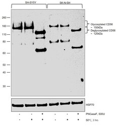
- Western blot was performed using Anti-CD56 Monoclonal Antibody (123C3) (Product # MA5-16446) and a 150 kDa band corresponding to CD56 was observed in SH-SY5Y and SK-N-SH. Membrane enriched cell extracts (12.5 µg lysate) of SH-SY5Y-Untreated (Lane 1), SH-SY5Y-Control (50°C, 3 hrs) (Lane 2), SH-SY5Y-Treated (500U PNGaseF; 50°C, 3 hr) (Lane 3), SK-N-SH-Untreated (Lane 4), SK-N-SH-Control (50°C, 3 hrs) (Lane 5) and SK-N-SH -Treated (500U PNGaseF; 50°C, 3 hr) (Lane 6) were electrophoresed using NuPAGE™ 4-12% Bis-Tris Protein Gel (Product # NP0321BOX). Resolved proteins were transferred onto a Nitrocellulose membrane (Product # IB23001) by iBlot® 2 Dry Blotting System (Product # IB21001). The blot was probed with the primary antibody (1:1,000 dilution) and detected by chemiluminescence with Goat anti-Mouse IgG (H+L) Superclonal™ Recombinant Secondary Antibody, HRP (Product # A28177, 1:20,000 dilution) using the iBright FL1500 (Product # A44115). Chemiluminescent detection was performed using SuperSignal™ West Dura Extended Duration Substrate (Product # 34076). CD56 migrates at ~150 kDa due to multiple glycosylations (Lane 1 and Lane 4). To carry out deglycosylation of the protein, PNGase F Glycan Cleavage Kit was used (Product # A39245). Upon PNGaseF treatment, N-linked glycosylations are cleaved and deglycosylated CD56 can be seen at ~120 kDa (Lane 3 and Lane 6). This mass shift can be attributed to deglycosylation as the control sample

- Submitted by
- Invitrogen Antibodies (provider)
- Main image
- Experimental details
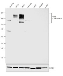
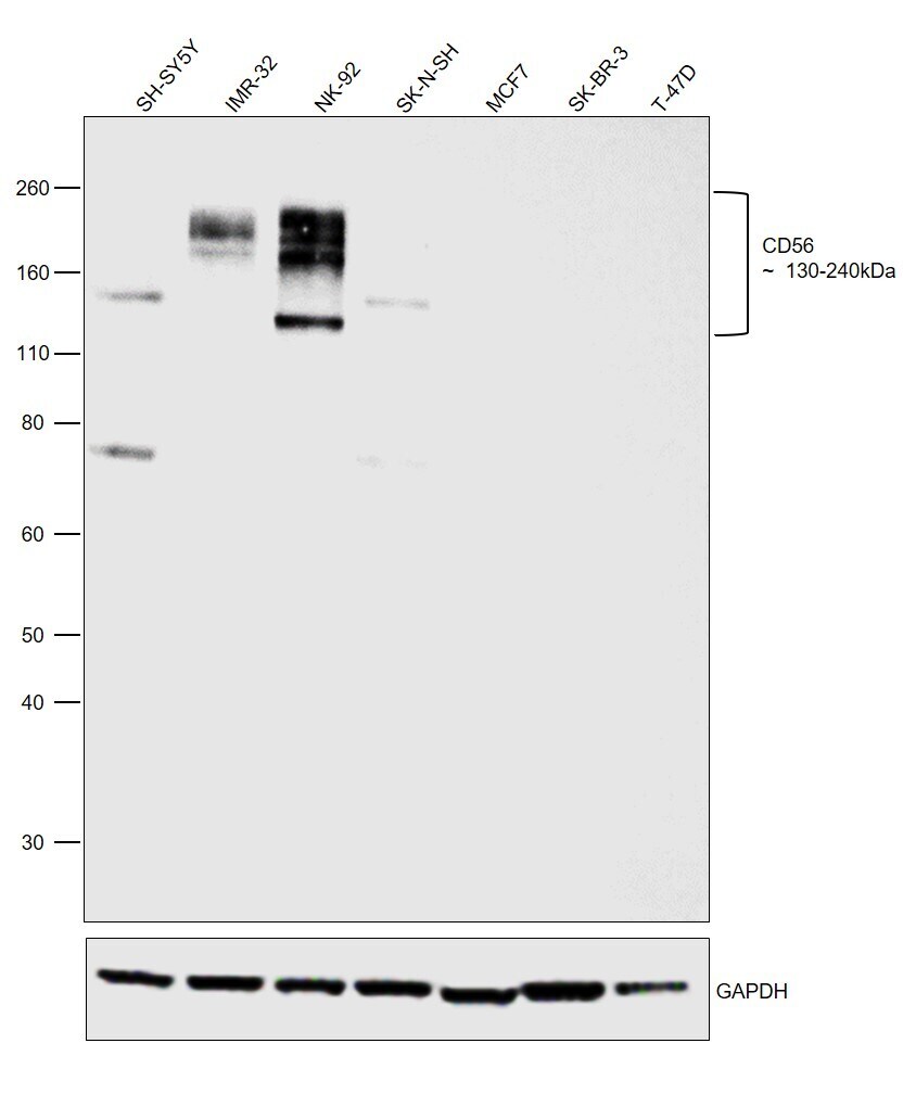
- Western blot was performed using Anti-CD56 Monoclonal Antibody (123C3) (Product # MA5-16445, MA5-16446) and bands in the range of 130-240kDa corresponding to Neural cell adhesion molecule 1 were observed.. Membrane enriched extracts (30 µg lysate) of SH-SY5Y (Lane 1), IMR-32 (Lane 2), NK-92 (Lane 3), SK-N-SH (Lane 4), MCF7 (Lane 5), SK-BR-3 (Lane 6) and T-47D (Lane 7) were electrophoresed using NuPAGE™ 4-12% Bis-Tris Protein Gel (Product # NP0322BOX). Resolved proteins were then transferred onto a nitrocellulose membrane (Product # IB23001) by iBlot® 2 Dry Blotting System (Product # IB21001). The blot was probed with the primary antibody (1:1000) and detected by chemiluminescence with Goat anti-Mouse IgG (H+L) Superclonal™ Recombinant Secondary Antibody, HRP (Product # A28177,1:20,000 using the iBright™ FL1500 Imaging System (Product # A44115). Chemiluminescent detection was performed using SuperSignal™ West Dura Extended Duration Substrate (Product # 34076). Relative expression was observed between SH-SY5Y, IMR-32, NK-92, SK-N-SH and breast cancer cell lines such as MCF7, SK-BR-3 and T-47D as expected (DOI: 10.1038/s41598-019-45377-8).
Supportive validation
- Submitted by
- Invitrogen Antibodies (provider)
- Main image
- Experimental details
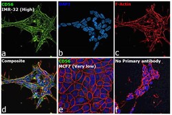
- Immunofluorescence analysis of Neural cell adhesion molecule 1 was performed using 70% confluent log phase IMR-32 cells. The cells were fixed with 4% paraformaldehyde for 10 minutes and blocked with 2% BSA for 1 hour at room temperature. The cells were labeled with CD56 Monoclonal Antibody (123C3) (Product # MA5-16445, MA5-16446) at 1:200 in 0.1% BSA, incubated at 4 degree celsius overnight and then labeled with Donkey anti-Mouse IgG (H+L) Highly Cross-Adsorbed Secondary Antibody, Alexa Fluor Plus 488 (Product # A32766), (1:2000), for 45 minutes at room temperature (Panel a: Green). Nuclei (Panel b:Blue) were stained with ProLong™ Diamond Antifade Mountant with DAPI (Product # P36962). F-actin (Panel c: Red) was stained with Rhodamine Phalloidin (Product # R415, 1:300). Panel d represents the merged image showing Plasma membrane localization. Panel e represents the merged image of MCF7 cells having very low to nil expression of CD56 as expected (DOI: 10.1038/s41598-019-45377-8). Panel f represents control cells with no primary antibody to assess background. The images were captured at 60X magnification.
