Explore
 Validate
 Learn
Learn Western blot
 Immunocytochemistry
Antibody data
- Antibody Data
- Antigen structure
- References [5]
- Comments [0]
- Validations
- Immunocytochemistry [1]
- Immunohistochemistry [1]
Submit
Validation data
Reference
Comment
Report error
- Product number
- HPA039835 - Provider product page

- Provider
- Atlas Antibodies
- Proper citation
- Atlas Antibodies Cat#HPA039835, RRID:AB_2676705
- Product name
- Anti-NCAM1
- Antibody type
- Polyclonal
- Description
- Polyclonal Antibody against Human NCAM1, Gene description: neural cell adhesion molecule 1, Alternative Gene Names: CD56, NCAM, Validated applications: IHC, WB, ICC, Uniprot ID: P13591, Storage: Store at +4°C for short term storage. Long time storage is recommended at -20°C.
- Reactivity
- Human
- Host
- Rabbit
- Conjugate
- Unconjugated
- Isotype
- IgG
- Vial size
- 100 µl
- Concentration
- 0.1 mg/ml
- Storage
- Store at +4°C for short term storage. Long time storage is recommended at -20°C.
- Handling
- The antibody solution should be gently mixed before use.
Submitted references Clinical and Histopathologic Characteristics of Pediatric Patients With Primary Membranous Nephropathy.
Discovery of seven novel putative antigens in membranous nephropathy and membranous lupus nephritis identified by mass spectrometry.
Neural cell adhesion molecule 1 is a novel autoantigen in membranous lupus nephritis.
Transforming Growth Factor Beta Receptor 3 (TGFBR3)-Associated Membranous Nephropathy.
Clinicoradiological characteristics, management and prognosis of primary myeloid sarcoma of the central nervous system: A report of four cases
Kouri AM, Caza TN, Beck LH Jr, Misurac JM, Evans MD, Phillips CL, Eadon MT, Larsen CP, Andreoli SP, Bu L, Rheault MN, Khalid M
Kidney international reports 2023 Nov;8(11):2368-2375
Kidney international reports 2023 Nov;8(11):2368-2375
Discovery of seven novel putative antigens in membranous nephropathy and membranous lupus nephritis identified by mass spectrometry.
Caza TN, Storey AJ, Hassen SI, Herzog C, Edmondson RD, Arthur JM, Kenan DJ, Larsen CP
Kidney international 2023 Mar;103(3):593-606
Kidney international 2023 Mar;103(3):593-606
Neural cell adhesion molecule 1 is a novel autoantigen in membranous lupus nephritis.
Caza TN, Hassen SI, Kuperman M, Sharma SG, Dvanajscak Z, Arthur J, Edmondson R, Storey A, Herzog C, Kenan DJ, Larsen CP
Kidney international 2021 Jul;100(1):171-181
Kidney international 2021 Jul;100(1):171-181
Transforming Growth Factor Beta Receptor 3 (TGFBR3)-Associated Membranous Nephropathy.
Caza TN, Hassen SI, Kenan DJ, Storey A, Arthur JM, Herzog C, Edmondson RD, Bourne TD, Beck LH Jr, Larsen CP
Kidney360 2021 Aug 26;2(8):1275-1286
Kidney360 2021 Aug 26;2(8):1275-1286
Clinicoradiological characteristics, management and prognosis of primary myeloid sarcoma of the central nervous system: A report of four cases
Yang B, Yang C, Fang J, Yang J, Xu Y
Oncology Letters 2017;14(3):3825-3831
Oncology Letters 2017;14(3):3825-3831
No comments: Submit comment
Supportive validation
- Submitted by
- Atlas Antibodies (provider)
- Main image
- Experimental details
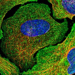
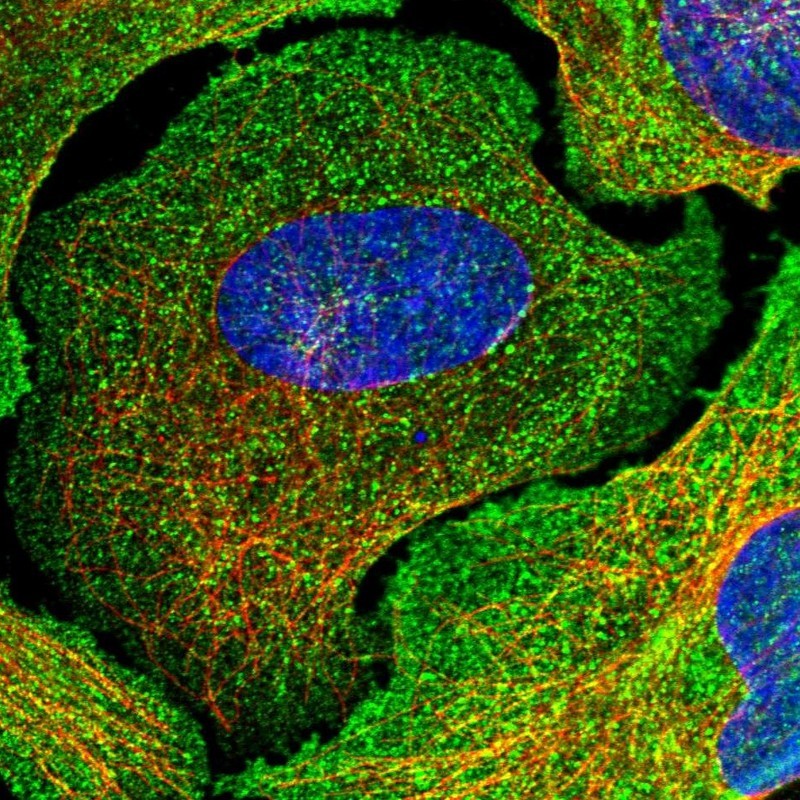
- Immunofluorescent staining of human cell line U-2 OS shows localization to plasma membrane & cytosol.
- Sample type
- Human
Supportive validation
- Submitted by
- Atlas Antibodies (provider)
- Enhanced method
- Orthogonal validation
- Main image
- Experimental details
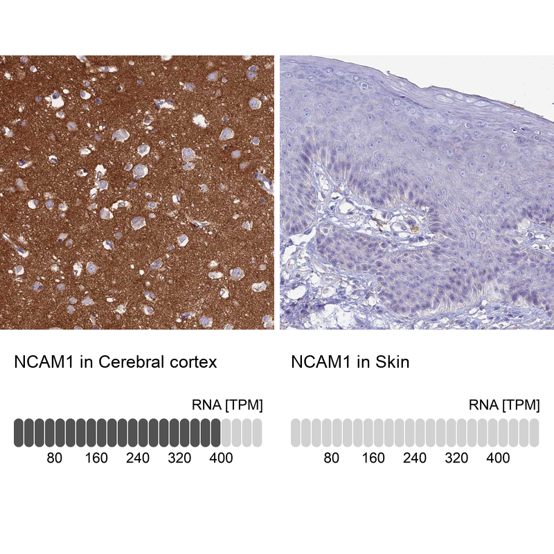
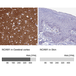
- Immunohistochemistry analysis in human cerebral cortex and skin tissues using HPA039835 antibody. Corresponding NCAM1 RNA-seq data are presented for the same tissues.
- Sample type
- Human
- Protocol
- Protocol