Explore
 Validate
 Learn
Learn Western blot
 Immunoprecipitation
 Immunohistochemistry
Antibody data
- Antibody Data
- Antigen structure
- References [21]
- Comments [0]
- Validations
- Immunohistochemistry [3]
- Other assay [5]
Submit
Validation data
Reference
Comment
Report error
- Product number
- 07-5603 - Provider product page

- Provider
- Invitrogen Antibodies
- Product name
- CD56 Monoclonal Antibody (123C3)
- Antibody type
- Monoclonal
- Antigen
- Other
- Reactivity
- Human
- Host
- Mouse
- Isotype
- IgG
- Antibody clone number
- 123C3
- Vial size
- 100 μg
- Concentration
- 1 mg/mL
- Storage
- -20°C
Submitted references An omic and multidimensional spatial atlas from serial biopsies of an evolving metastatic breast cancer.
The prognostic significance of tumor-infiltrating lymphocytes in cervical cancer.
Deep spatial profiling of human COVID-19 brains reveals neuroinflammation with distinct microanatomical microglia-T-cell interactions.
Parvovirus B19-Infected Tubulointerstitial Nephritis in Hereditary Spherocytosis.
Usefulness of the octreotide test in Japanese patients for predicting the presence/absence of somatostatin receptor 2 expression in insulinomas.
Variable Expression of Neural Cell Adhesion Molecule Isoforms in Renal Tissue: Possible Role in Incipient Renal Fibrosis.
Canalicular adenoma: a clinicopathologic and immunohistochemical analysis of 67 cases with a review of the literature.
Arterial flow regulator enables transplantation and growth of human fetal kidneys in rats.
Neuronal Subtype and Satellite Cell Tropism Are Determinants of Varicella-Zoster Virus Virulence in Human Dorsal Root Ganglia Xenografts In Vivo.
A case of (123)I-MIBG scintigram-negative functioning pheochromocytoma: immunohistochemical and molecular analysis with review of literature.
CD30(+) extranodal natural killer/T-cell lymphoma mimicking phlegmonous myositis: A case report.
A Case of Distal Epithelioid Sarcoma of the Thumb Expressing Podoplanin, TLE1 and Ca 125.
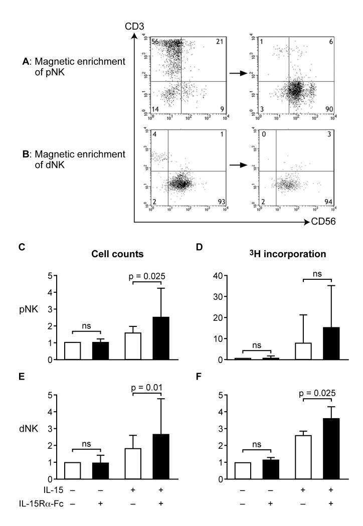
Uterine NK cells regulate endometrial bleeding in women and are suppressed by the progesterone receptor modulator asoprisnil.
Serous effusion cytology of extranodal natural killer/T-cell lymphoma.
Extraosseous (extramedullary) plasmacytomas: a clinicopathologic and immunophenotypic study of 32 Chinese cases.
Subcutaneous panniculitis-like T-cell lymphoma: a clinical and pathologic study of 14 korean patients.
Goblet cell carcinoid of the appendix: a clinicopathological and immunohistochemical study of 26 cases from southwest china.
Varicella-zoster virus infection of human neural cells in vivo.
Simian virus 40 sequences in malignant lymphomas in Japan.
Evasion from NK cell immunity by MHC class I chain-related molecules expressing colon adenocarcinoma.
Evasion from NK cell immunity by MHC class I chain-related molecules expressing colon adenocarcinoma.
Johnson BE, Creason AL, Stommel JM, Keck JM, Parmar S, Betts CB, Blucher A, Boniface C, Bucher E, Burlingame E, Camp T, Chin K, Eng J, Estabrook J, Feiler HS, Heskett MB, Hu Z, Kolodzie A, Kong BL, Labrie M, Lee J, Leyshock P, Mitri S, Patterson J, Riesterer JL, Sivagnanam S, Somers J, Sudar D, Thibault G, Weeder BR, Zheng C, Nan X, Thompson RF, Heiser LM, Spellman PT, Thomas G, Demir E, Chang YH, Coussens LM, Guimaraes AR, Corless C, Goecks J, Bergan R, Mitri Z, Mills GB, Gray JW
Cell reports. Medicine 2022 Feb 15;3(2):100525
Cell reports. Medicine 2022 Feb 15;3(2):100525
The prognostic significance of tumor-infiltrating lymphocytes in cervical cancer.
He M, Wang Y, Zhang G, Cao K, Yang M, Liu H
Journal of gynecologic oncology 2021 May;32(3):e32
Journal of gynecologic oncology 2021 May;32(3):e32
Deep spatial profiling of human COVID-19 brains reveals neuroinflammation with distinct microanatomical microglia-T-cell interactions.
Schwabenland M, Salié H, Tanevski J, Killmer S, Lago MS, Schlaak AE, Mayer L, Matschke J, Püschel K, Fitzek A, Ondruschka B, Mei HE, Boettler T, Neumann-Haefelin C, Hofmann M, Breithaupt A, Genc N, Stadelmann C, Saez-Rodriguez J, Bronsert P, Knobeloch KP, Blank T, Thimme R, Glatzel M, Prinz M, Bengsch B
Immunity 2021 Jul 13;54(7):1594-1610.e11
Immunity 2021 Jul 13;54(7):1594-1610.e11
Parvovirus B19-Infected Tubulointerstitial Nephritis in Hereditary Spherocytosis.
Nishiyama K, Watanabe Y, Ishimura M, Tetsuhara K, Imai T, Kanemasa H, Ueki K, Motomura Y, Kaku N, Sakai Y, Imadome KI, Ohga S
Open forum infectious diseases 2020 Aug;7(8):ofaa288
Open forum infectious diseases 2020 Aug;7(8):ofaa288
Usefulness of the octreotide test in Japanese patients for predicting the presence/absence of somatostatin receptor 2 expression in insulinomas.
Nakamura A, Mitsuhashi T, Takano Y, Miyoshi H, Kameda H, Nomoto H, Nagai S, Hatanaka Y, Shimizu C, Terauchi Y, Atsumi T
Endocrine journal 2016;63(2):135-42
Endocrine journal 2016;63(2):135-42
Variable Expression of Neural Cell Adhesion Molecule Isoforms in Renal Tissue: Possible Role in Incipient Renal Fibrosis.
Marković-Lipkovski J, Životić M, Müller CA, Tampe B, Ćirović S, Vještica J, Tomanović N, Zeisberg M, Müller GA
PloS one 2015;10(9):e0137028
PloS one 2015;10(9):e0137028
Canalicular adenoma: a clinicopathologic and immunohistochemical analysis of 67 cases with a review of the literature.
Thompson LD, Bauer JL, Chiosea S, McHugh JB, Seethala RR, Miettinen M, Müller S
Head and neck pathology 2015 Jun;9(2):181-95
Head and neck pathology 2015 Jun;9(2):181-95
Arterial flow regulator enables transplantation and growth of human fetal kidneys in rats.
Chang NK, Gu J, Gu S, Osorio RW, Concepcion W, Gu E
American journal of transplantation : official journal of the American Society of Transplantation and the American Society of Transplant Surgeons 2015 Jun;15(6):1692-700
American journal of transplantation : official journal of the American Society of Transplantation and the American Society of Transplant Surgeons 2015 Jun;15(6):1692-700
Neuronal Subtype and Satellite Cell Tropism Are Determinants of Varicella-Zoster Virus Virulence in Human Dorsal Root Ganglia Xenografts In Vivo.
Zerboni L, Arvin A
PLoS pathogens 2015 Jun;11(6):e1004989
PLoS pathogens 2015 Jun;11(6):e1004989
A case of (123)I-MIBG scintigram-negative functioning pheochromocytoma: immunohistochemical and molecular analysis with review of literature.
Kurisaki-Arakawa A, Saito T, Takahashi M, Mitani K, Yao T
International journal of clinical and experimental pathology 2014;7(7):4438-47
International journal of clinical and experimental pathology 2014;7(7):4438-47
CD30(+) extranodal natural killer/T-cell lymphoma mimicking phlegmonous myositis: A case report.
Yang YJ, Li YX, Liu YB, Yang M, Liu K
Oncology letters 2014 May;7(5):1419-1421
Oncology letters 2014 May;7(5):1419-1421
A Case of Distal Epithelioid Sarcoma of the Thumb Expressing Podoplanin, TLE1 and Ca 125.
Karagkounis G, Argyrakos T, Charkiolakis G, Castana O, Rontogianni D
Case reports in pathology 2013;2013:312786
Case reports in pathology 2013;2013:312786
Uterine NK cells regulate endometrial bleeding in women and are suppressed by the progesterone receptor modulator asoprisnil.
Wilkens J, Male V, Ghazal P, Forster T, Gibson DA, Williams AR, Brito-Mutunayagam SL, Craigon M, Lourenco P, Cameron IT, Chwalisz K, Moffett A, Critchley HO
Journal of immunology (Baltimore, Md. : 1950) 2013 Sep 1;191(5):2226-35
Journal of immunology (Baltimore, Md. : 1950) 2013 Sep 1;191(5):2226-35
Serous effusion cytology of extranodal natural killer/T-cell lymphoma.
Su XY, Huang J, Jiang Y, Tang Y, Li GD, Liu WP
Cytopathology : official journal of the British Society for Clinical Cytology 2012 Apr;23(2):96-102
Cytopathology : official journal of the British Society for Clinical Cytology 2012 Apr;23(2):96-102
Extraosseous (extramedullary) plasmacytomas: a clinicopathologic and immunophenotypic study of 32 Chinese cases.
Zuo Z, Tang Y, Bi CF, Zhang WY, Zhao S, Wang XQ, Yang QP, Zou LQ, Liu WP
Diagnostic pathology 2011 Dec 19;6:123
Diagnostic pathology 2011 Dec 19;6:123
Subcutaneous panniculitis-like T-cell lymphoma: a clinical and pathologic study of 14 korean patients.
Lee DW, Yang JH, Lee SM, Won CH, Chang S, Lee MW, Choi JH, Moon KC
Annals of dermatology 2011 Aug;23(3):329-37
Annals of dermatology 2011 Aug;23(3):329-37
Goblet cell carcinoid of the appendix: a clinicopathological and immunohistochemical study of 26 cases from southwest china.
Yong Jiang, Huawei Liu, Hu Long, Yingying Yang, Dianying Liao, Xiuhui Zhang
International journal of surgical pathology 2010 Dec;18(6):488-92
International journal of surgical pathology 2010 Dec;18(6):488-92
Varicella-zoster virus infection of human neural cells in vivo.
Baiker A, Fabel K, Cozzio A, Zerboni L, Fabel K, Sommer M, Uchida N, He D, Weissman I, Arvin AM
Proceedings of the National Academy of Sciences of the United States of America 2004 Jul 20;101(29):10792-7
Proceedings of the National Academy of Sciences of the United States of America 2004 Jul 20;101(29):10792-7
Simian virus 40 sequences in malignant lymphomas in Japan.
Nakatsuka S, Liu A, Dong Z, Nomura S, Takakuwa T, Miyazato H, Aozasa K, Osaka Lymphoma Study Group
Cancer research 2003 Nov 15;63(22):7606-8
Cancer research 2003 Nov 15;63(22):7606-8
Evasion from NK cell immunity by MHC class I chain-related molecules expressing colon adenocarcinoma.
Doubrovina ES, Doubrovin MM, Vider E, Sisson RB, O'Reilly RJ, Dupont B, Vyas YM
Journal of immunology (Baltimore, Md. : 1950) 2003 Dec 15;171(12):6891-9
Journal of immunology (Baltimore, Md. : 1950) 2003 Dec 15;171(12):6891-9
No comments: Submit comment
Supportive validation
- Submitted by
- Invitrogen Antibodies (provider)
- Main image
- Experimental details
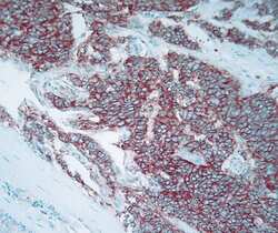
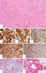
- Immunohistochemical staining of small cell lung cancer tissue using Mouse anti-CD56 monoclonal antibody (clone 123C3) (Product # 07-5603).
- Submitted by
- Invitrogen Antibodies (provider)
- Main image
- Experimental details
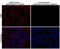
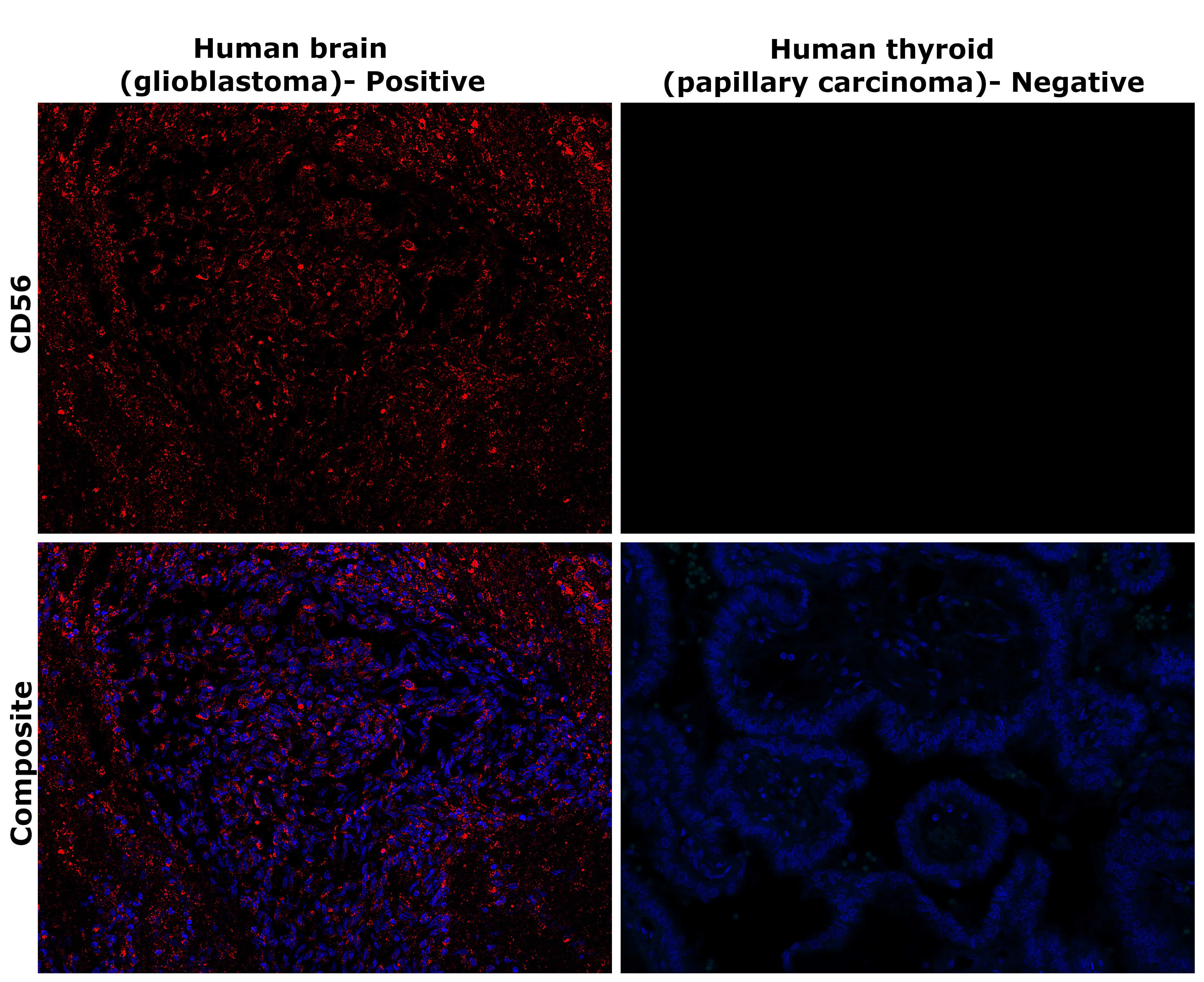
- Immunohistochemical analysis of CD56 (NCAM) was performed using formalin-fixed paraffin-embedded human brain (glioblastoma) and human thyroid (thyroid- profiling papillary carcinoma) tissue sections. To expose the target protein, heat-induced epitope retrieval was performed on de-paraffinized sections using eBioscience™ IHC Antigen Retrieval Solution - Low pH (10X) (Product # 00-4955-58) diluted to 1X solution in water in a decloaking chamber at 110 degree Celsius for 15 minutes. Following antigen retrieval, the sections were blocked with 3% H2O2 for 1 hour at room temperature followed by 2% normal goat serum in 1X PBS for 45 minutes at room temperature and then probed with CD56 Monoclonal Antibody (123C3) (Product # 07-5603) at 2 µg/mL in 0.1% normal goat serum overnight at 4 degree Celsius in a humidified chamber. Detection was performed using Alexa Fluor™ 647 Tyramide SuperBoost™ Kit, goat anti-mouse IgG (Product # B40916). Nuclei were stained with DAPI (Product # D1306) and the sections were mounted using ProLong™ Glass Antifade Mountant (Product # P36984). The images were captured on EVOS™ M7000 Imaging System (Product # AMF7000) at 20X magnification and externally deconvoluted.
- Submitted by
- Invitrogen Antibodies (provider)
- Main image
- Experimental details
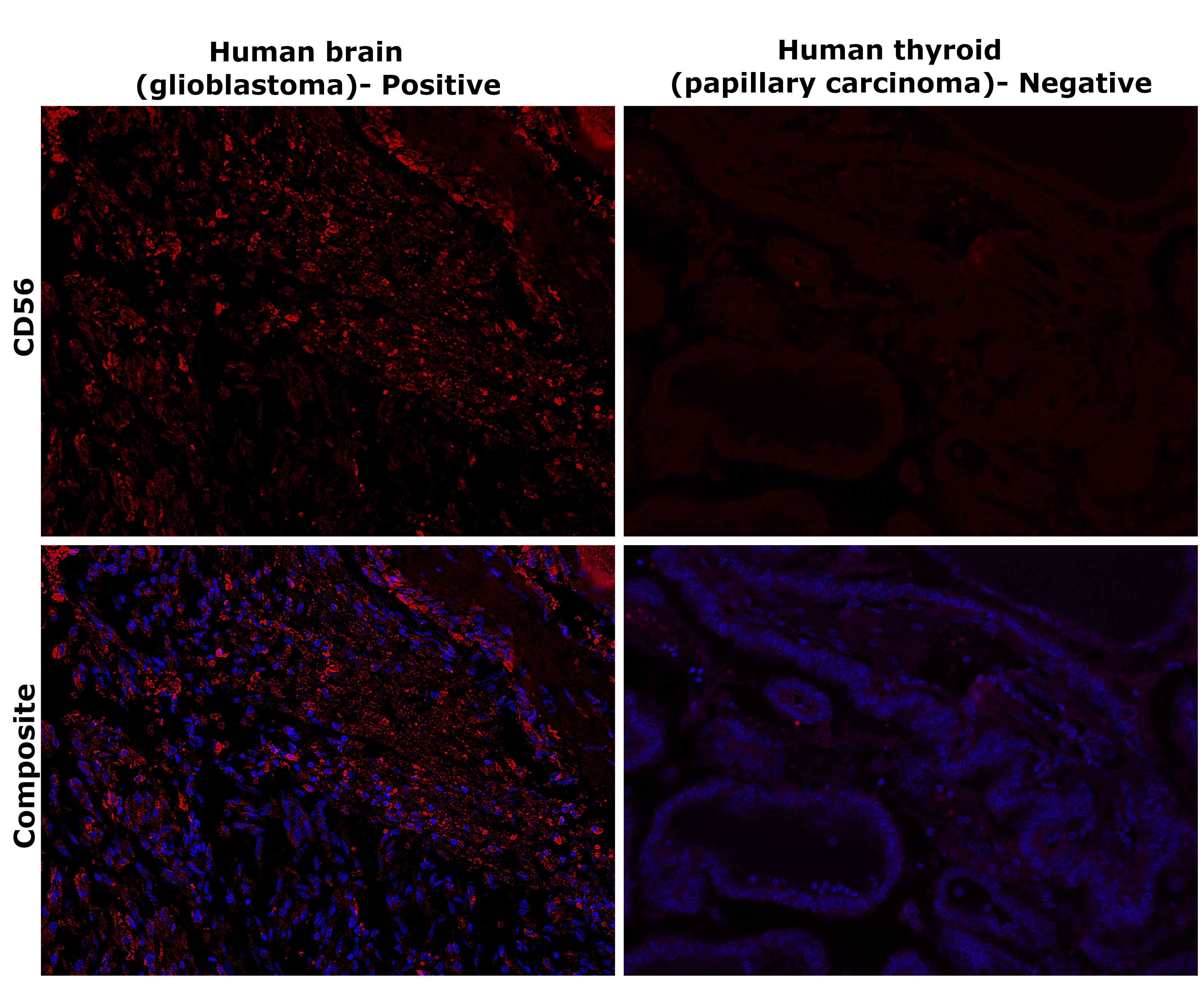
- Immunohistochemical analysis of CD56 (NCAM) was performed using formalin-fixed paraffin-embedded human brain (glioblastoma) and human thyroid (thyroid- profiling papillary carcinoma) tissue sections. To expose the target protein, heat-induced epitope retrieval was performed on de-paraffinized sections using eBioscience™ IHC Antigen Retrieval Solution - Low pH (10X) (Product # 00-4955-58) diluted to 1X solution in water in a decloaking chamber at 110 degree Celsius for 15 minutes. Following antigen retrieval, the sections were blocked with 3% H2O2 for 1 hour at room temperature followed by 2% normal goat serum in 1X PBS for 45 minutes at room temperature and then probed with CD56 Monoclonal Antibody (123C3) (Product # 07-5603) at 2 µg/mL in 0.1% normal goat serum overnight at 4 degree Celsius in a humidified chamber. Detection was performed using Alexa Fluor™ 647 Tyramide SuperBoost™ Kit, goat anti-mouse IgG (Product # B40916). Nuclei were stained with DAPI (Product # D1306) and the sections were mounted using ProLong™ Glass Antifade Mountant (Product # P36984). The images were captured on EVOS™ M7000 Imaging System (Product # AMF7000) at 20X magnification and externally deconvoluted.
Supportive validation
- Submitted by
- Invitrogen Antibodies (provider)
- Main image

- Experimental details
- NULL
- Submitted by
- Invitrogen Antibodies (provider)
- Main image
- Experimental details
- NULL

- Submitted by
- Invitrogen Antibodies (provider)
- Main image
- Experimental details
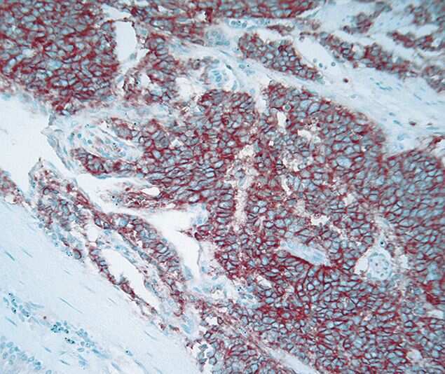
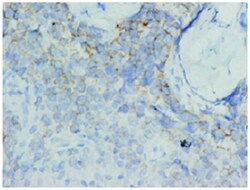
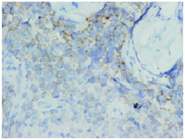
- Figure 3 Immunohistochemistry revealed that the majority of tumor cells were positive CD56 staining in portions (3,3'-diaminobenzidine staining magnification, x500).
- Submitted by
- Invitrogen Antibodies (provider)
- Main image
- Experimental details
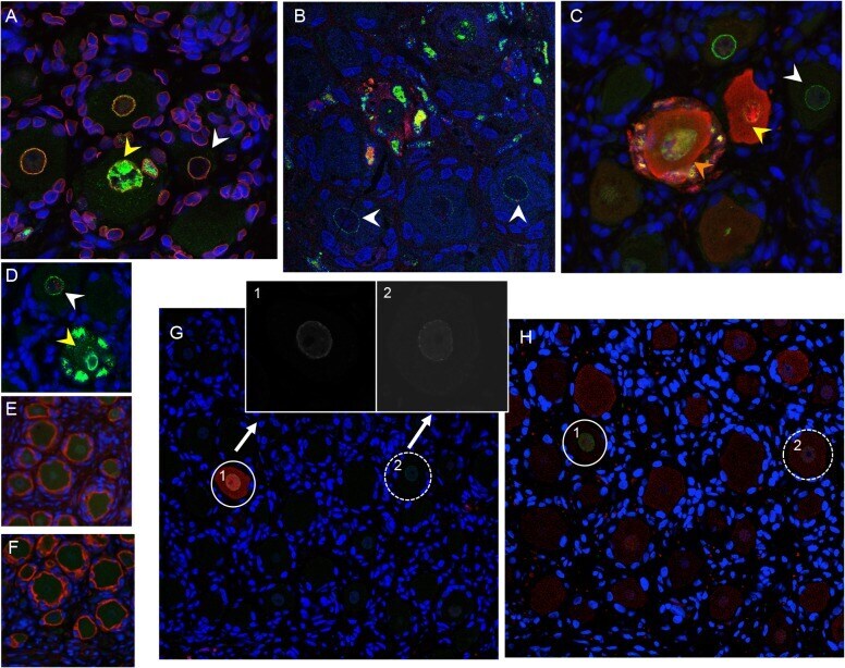
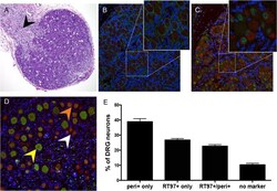
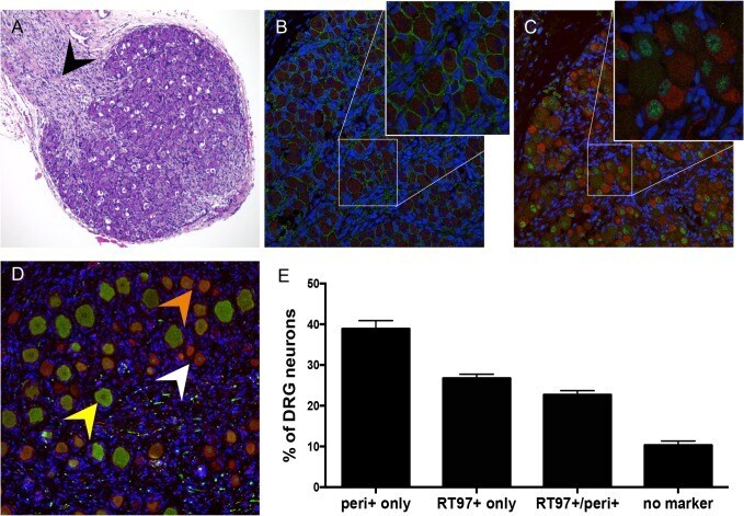
- Fig 1 Differentiation of neurons after DRG implantation. Panels A-C, fetal DRG, 18 gestational weeks, prior to subcapsular renal implantation in SCID mouse, stained with (A) hematoxylin & eosin; black arrow denotes nerve root, (B) anti-NCAM (green)/anti-synaptophysin (red) antibody, and (C) subtype specific markers anti-RT97 (green)/anti-peripherin (red) antibody. Panels D-E, DRG xenograft, 20 weeks after transplantation, stained with subtype specific markers anti-RT97 (green)/anti-peripherin (red) antibody (D), demonstrates that neuronal differentiation continues in DRG xengrafts in vivo . Peripherin staining (white arrow) identifies nociceptive neurons; cytoplasmic RT97 immunoreactivity (yellow arrow) identifies mechanoreceptive neurons. Cell counting (E) determined the proportion of nociceptive and RT97-immunoreactive mechanoreceptive neurons at 20 weeks after implantation. For each panel, representative images are shown. For assessment of neuronal subtype at least 20 fields were counted, each with a minimum of 50 neurons.
- Submitted by
- Invitrogen Antibodies (provider)
- Main image
- Experimental details
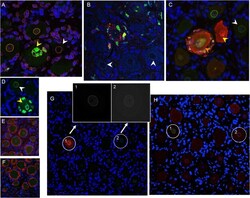
- Fig 5 VZV restriction in mechanoreceptive neurons occurs after viral entry. For each panel, one representative image is shown for staining experiments performed on multiple tissue sections (6-10 sections) for each DRG. (A-C) ORF23 capsid protein is a marker of virion entry as well as the later formation of progeny virions; virion entry is indicated by ORF23 capsid puncta at the nuclear rim (green), co-stained with cellular lamin A/C (A, red), and prior to expression of IE62 (B, red) or IE63 (C, red). (D) Dual staining for ORF23 (green) and cellular PML (red). (E) ORF23 nuclear rim staining was not observed when using pre-immune serum (green); red is N-CAM staining. (F) ORF23 capsid protein is not detected in uninfected DRG, as shown by staining for N-CAM (red) and absence of ORF23 (green). (G-H) Staining of adjacent tissue sections stained with antibody for ORF23 (green) and IE63 (G, red) or RT97 (H, red). Two neurons are circled and shown in both panels. The single 488-channel images for Panel G, shown in greyscale for better visualization, are provided for the circled neurons (1 and 2). The contrast of the G, neuron 2, ORF23 B&W panel, is enhanced so that the discrete ORF23 particles are easier to observe.