Explore
 Validate
 Learn
Learn Western blot
 Flow cytometry
Antibody data
- Antibody Data
- Antigen structure
- References [2]
- Comments [0]
- Validations
- Western blot [2]
- Immunocytochemistry [2]
Submit
Validation data
Reference
Comment
Report error
- Product number
- AF2408 - Provider product page

- Provider
- R&D Systems
- Product name
- Human/Mouse NCAM-1/CD56 Antibody
- Antibody type
- Polyclonal
- Description
- Antigen Affinity-purified. Detects human NCAM-1/CD56 in direct ELISAs and Western blots. In direct ELISAs and Western blots, less than 1% cross-reactivity with recombinant human (rh) ALCAM, rhBCAM and rhEpCAM is observed.
- Reactivity
- Human, Mouse
- Host
- Goat
- Conjugate
- Unconjugated
- Antigen sequence
NP_001070150- Isotype
- IgG
- Vial size
- 100 ug
- Concentration
- LYOPH
- Storage
- Use a manual defrost freezer and avoid repeated freeze-thaw cycles. 12 months from date of receipt, -20 to -70 °C as supplied. 1 month, 2 to 8 °C under sterile conditions after reconstitution. 6 months, -20 to -70 °C under sterile conditions after reconstitution.
Submitted references Modeling Developmental and Tumorigenic Aspects of Trilateral Retinoblastoma via Human Embryonic Stem Cells.
Divergent modulation of neuronal differentiation by caspase-2 and -9.
Avior Y, Lezmi E, Yanuka D, Benvenisty N
Stem cell reports 2017 May 9;8(5):1354-1365
Stem cell reports 2017 May 9;8(5):1354-1365
Divergent modulation of neuronal differentiation by caspase-2 and -9.
Pistritto G, Papaleo V, Sanchez P, Ceci C, Barbaccia ML
PloS one 2012;7(5):e36002
PloS one 2012;7(5):e36002
No comments: Submit comment
Supportive validation
- Submitted by
- R&D Systems (provider)
- Main image
- Experimental details
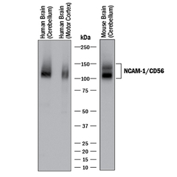
- Detection of Human and Mouse NCAM-1/CD56 by Western Blot. Western blot shows lysates of human brain (cerebellum and motor cortex) tissue and mouse brain (cerebellum) tissue. PVDF membrane was probed with 0.5 µg/mL of Goat Anti-Human/Mouse NCAM-1/CD56 Antigen Affinity-purified Polyclonal Antibody (Catalog # AF2408) followed by HRP-conjugated Anti-Goat IgG Secondary Antibody (Catalog # HAF017). Specific bands were detected for NCAM-1/CD56 at approximately 100-150 kDa (as indicated). This experiment was conducted under reducing conditions and using Immunoblot Buffer Group 1.
- Submitted by
- R&D Systems (provider)
- Main image
- Experimental details
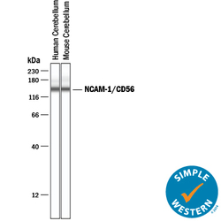
- Detection of Human and Mouse NCAM-1/CD56 by Simple WesternTM. Simple Western lane view shows lysates of human and mouse brain (cerebellum) tissue, loaded at 0.2 mg/mL. A specific band was detected for NCAM-1/CD56 at approximately 143 kDa (as indicated) using 5 µg/mL for human lysates and 25 µg/mL for mouse lysates of Goat Anti-Human/Mouse NCAM-1/ CD56 Antigen Affinity-purified Polyclonal Antibody (Catalog # AF2408) followed by 1:50 dilution of HRP-conjugated Anti-Goat IgG Secondary Antibody (Catalog # HAF109). This experiment was conducted under reducing conditions and using the 12-230 kDa separation system.

Supportive validation
- Submitted by
- R&D Systems (provider)
- Main image
- Experimental details
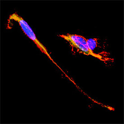
- NCAM-1/CD56 in SH-SY5Y Human Neuroblastoma Cells. SH-SY5Y human neuroblastoma cells were cultured overnight in the presence of 1 mM Retinoic Acid (Catalog # 0695/50) prior to immersion fixation. Neural Cell Adhesion Molecule 1 (NCAM-1)/CD56 was detected using a Goat Anti-Human/Mouse NCAM-1/CD56 Antigen Affinity-purified Polyclonal Antibody (Catalog # AF2408). The cells were stained with the NorthernLights 557-conjugated Donkey Anti-Goat IgG Affinity-purified Secondary Antibody (red; Catalog # NL001). Actin filaments were stained with FITC-conjugated Phalloidin (green) and cell nuclei were counter-stained with DAPI (blue). NCAM-1/CD56 immuno-reactivity was localized to the plasma membrane. View our protocol for Fluorescent ICC Staining of Cells on Coverslips.

- Submitted by
- R&D Systems (provider)
- Main image
- Experimental details
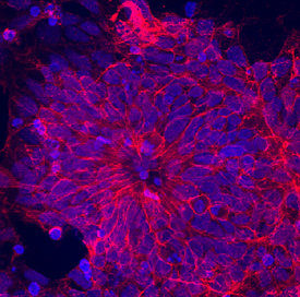
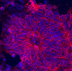
- NCAM-1/CD56 in BG01V Human Embryonic Stem Cells. NCAM-1/CD56 was detected in immersion fixed BG01V human embryonic stem cells differentiated into neural progenitor cells using Goat Anti-Human/Mouse NCAM-1/CD56 Antigen Affinity-purified Polyclonal Antibody (Catalog # AF2408) at 10 µg/mL for 3 hours at room temperature. Cells were stained using the NorthernLights™ 557-conjugated Anti-Goat IgG Secondary Antibody (red; Catalog # NL001) and counterstained with DAPI (blue). Specific staining was localized to cytoplasm. View our protocol for Fluorescent ICC Staining of Stem Cells on Coverslips.