Explore
 Validate
 Learn
Learn Western blot
 Immunohistochemistry
Antibody data
- Antibody Data
- Antigen structure
- References [1]
- Comments [0]
- Validations
- Immunohistochemistry [1]
- Other assay [2]
Submit
Validation data
Reference
Comment
Report error
- Product number
- PA5-34448 - Provider product page

- Provider
- Invitrogen Antibodies
- Product name
- FLT3 Polyclonal Antibody
- Antibody type
- Polyclonal
- Antigen
- Synthetic peptide
- Description
- A suggested positive control is NIH-3T3 cell lysate. PA5-34448 can be used with blocking peptide PEP-1490.
- Reactivity
- Human, Mouse
- Host
- Rabbit
- Isotype
- IgG
- Vial size
- 100 μg
- Concentration
- 1 mg/mL
- Storage
- Maintain refrigerated at 2-8°C for up to 3 months. For long term storage store at -20°C
Submitted references High-fat diet intensifies MLL-AF9-induced acute myeloid leukemia through activation of the FLT3 signaling in mouse primitive hematopoietic cells.
Hermetet F, Mshaik R, Simonet J, Callier P, Delva L, Quéré R
Scientific reports 2020 Sep 30;10(1):16187
Scientific reports 2020 Sep 30;10(1):16187
No comments: Submit comment
Supportive validation
- Submitted by
- Invitrogen Antibodies (provider)
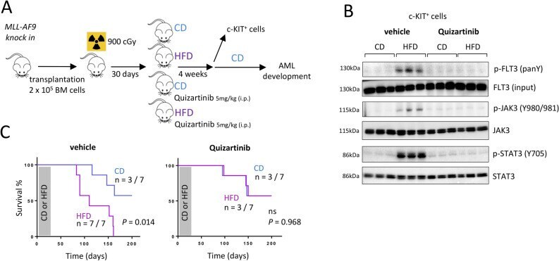
- Main image
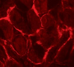
- Experimental details
- Immunofluorescence of FLT3 in human kidney tissue with FLT3 Polyclonal Antibody (Product # PA5-34448) at 20 µg/mL.
Supportive validation
- Submitted by
- Invitrogen Antibodies (provider)
- Main image

- Experimental details
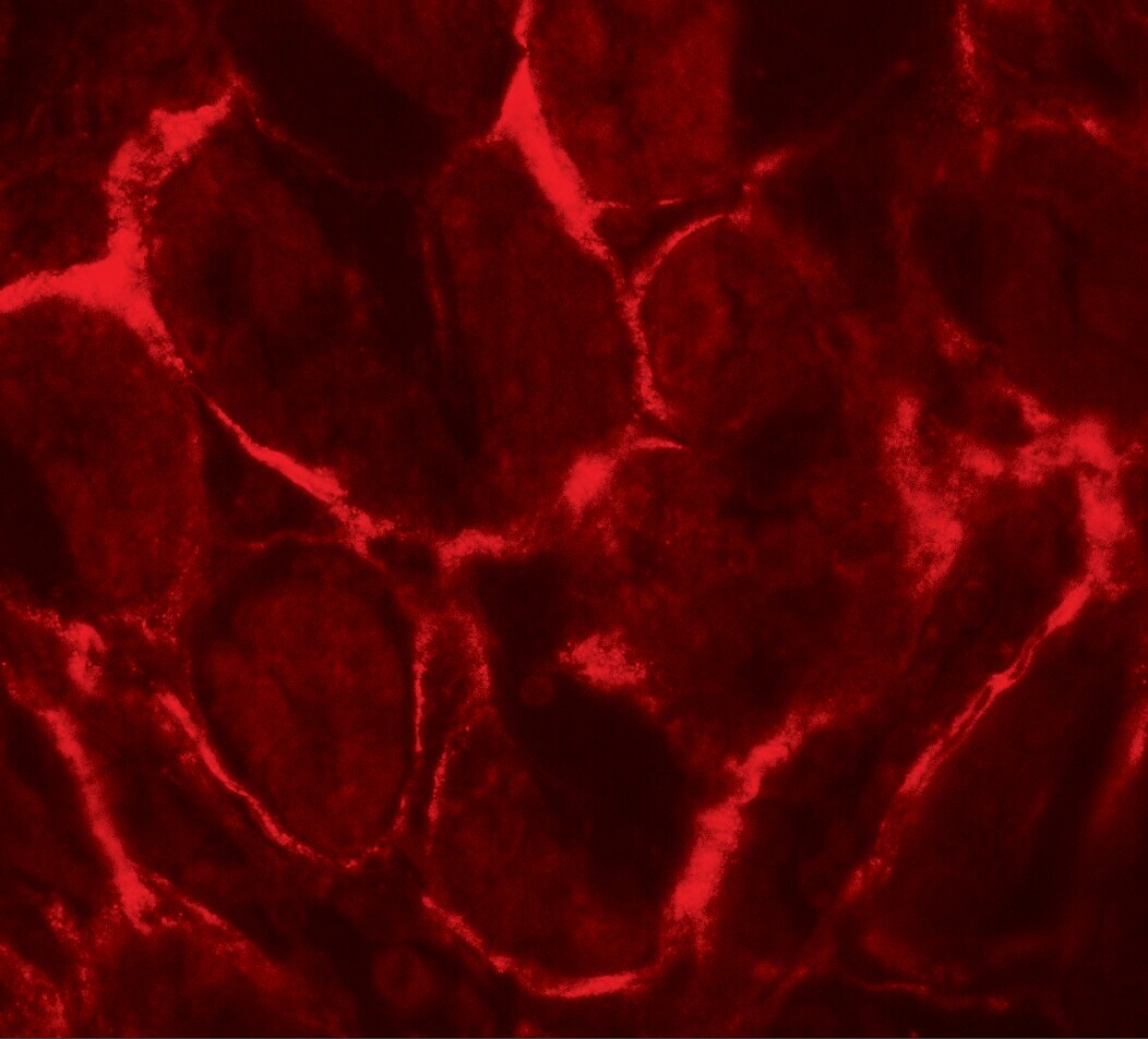
- Figure 3 HFD activates FLT3/JAK3/STAT3 signaling on c-KIT + BM cells after 4 weeks. ( A ) Western blot showing increased phosphorylation of JAK3 (Y980/981) and STAT3 (Y705) among the c-KIT + MLL-AF9 knock in cells in BM, after 4 weeks of HFD. Data show mean +- SD; n = 3 mice per diet group; *** P < 0.001, two-tailed unpaired Student''s t-test; ns, non-significant. Grouping of blots cropped from different gels, see Supplementary Fig. S6 for full-length blots. Gel images were processed and analyzed for quantification (Fiji, NIH). ( B ) Western blot showing increased phosphorylation of FLT3 among the c-KIT + MLL-AF9 knock in cells in BM, after 4-weeks of HFD. After immunoprecipitation of pan tyrosine phosphorylated proteins with an anti-phosphotyrosine antibody (IP: pan Y), FLT3 was analyzed by a western blot (WB: FLT3). Increased phosphorylation of FLT3 following HFD was confirmed by immunoprecipitation with an anti-FLT3 antibody (IP: FLT3) followed by a western blot to detect pan phosphorylation of FLT3 (WB: pan Y). Data show mean +- SD; n = 3 mice per diet group; *** P < 0.001, two-tailed unpaired Student''s t-test. Grouping of blots cropped from different gels, see Supplementary Fig. S7 for full-length blots. Gel images were processed and analyzed for quantification (Fiji, NIH). ( C ) Flow cytometry on MLL-AF9 knock in BM cells showing median fluorescence intensity (MFI) for expression of FLT3 on Lin - SCA1 + c-KIT + (LSK) or Lin - c-KIT + (LK) cells. Data were analyzed usin

- Submitted by
- Invitrogen Antibodies (provider)
- Main image
- Experimental details
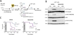
- Figure 4 Quizartinib blocks the HFD-accelerated development of AML. ( A ) Experimental workflow describing the procedure. Image performed with the GIMP software (version 2.10.18, GIMP, https://www.gimp.org/news/2020/02/24/gimp-2-10-18-released/ ). ( B ) Western blot showing that a treatment with Quizartinib antagonizes the increased phosphorylation of FLT3 (pan tyrosine; panY) as well as JAK3 (Y980/981) and STAT3 (Y705) observed among the c-KIT + MLL-AF9 knock in BM cells after 4-weeks of HFD. Data show mean +- SD; n = 3 mice per diet group. Grouping of blots cropped from different gels, see Supplementary Fig. S8 for full-length blots. Gel images were processed and analyzed for quantification (Fiji, NIH). ( C ) Survival curves showing that mice fed a HFD and treated with Quizartinib survived longer than CD-fed mice, n = 7 mice per group; P value measured by Mantel-Haenszel test.