 Explore
 Validate
 Learn
Learn Western blot
 Immunohistochemistry
Antibody data
- Antibody Data
- Antigen structure
- References [1]
- Comments [0]
- Validations
- Immunohistochemistry [4]
- Other assay [1]
Submit
Validation data
Reference
Comment
Report error
- Product number
- PA5-85901 - Provider product page

- Provider
- Invitrogen Antibodies
- Product name
- TMEM2 Polyclonal Antibody
- Antibody type
- Polyclonal
- Antigen
- Synthetic peptide
- Reactivity
- Human, Mouse, Rat
- Host
- Rabbit
- Isotype
- IgG
- Vial size
- 100 μL
- Concentration
- 1 mg/mL
- Storage
- Store at 4°C short term. For long term storage, store at -20°C, avoiding freeze/thaw cycles.
Submitted references Endogenously-Produced Hyaluronan and Its Potential to Regulate the Development of Peritoneal Adhesions.
Kocurkova A, Nesporova K, Sandanusova M, Kerberova M, Lehka K, Velebny V, Kubala L, Ambrozova G
Biomolecules 2021 Dec 29;12(1)
Biomolecules 2021 Dec 29;12(1)
No comments: Submit comment
Supportive validation
- Submitted by
- Invitrogen Antibodies (provider)
- Main image

- Experimental details
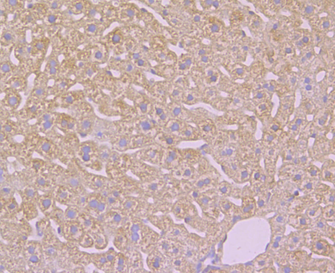
- Immunohistochemical analysis of TMEM2 of paraffin-embedded rat kidney tissue using a TMEM2 Polyclonal antibody (Product #PA5-85901). Counter stained with hematoxylin..
- Submitted by
- Invitrogen Antibodies (provider)
- Main image
- Experimental details
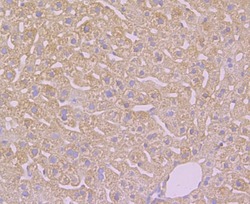
- Immunohistochemical analysis of TMEM2 of paraffin-embedded Human liver tissue using a TMEM2 Polyclonal antibody (Product #PA5-85901). Counter stained with hematoxylin..

- Submitted by
- Invitrogen Antibodies (provider)
- Main image
- Experimental details
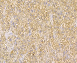
- Immunohistochemical analysis of TMEM2 of paraffin-embedded Mouse liver tissue using a TMEM2 Polyclonal antibody (Product #PA5-85901). Counter stained with hematoxylin..
- Submitted by
- Invitrogen Antibodies (provider)
- Main image
- Experimental details
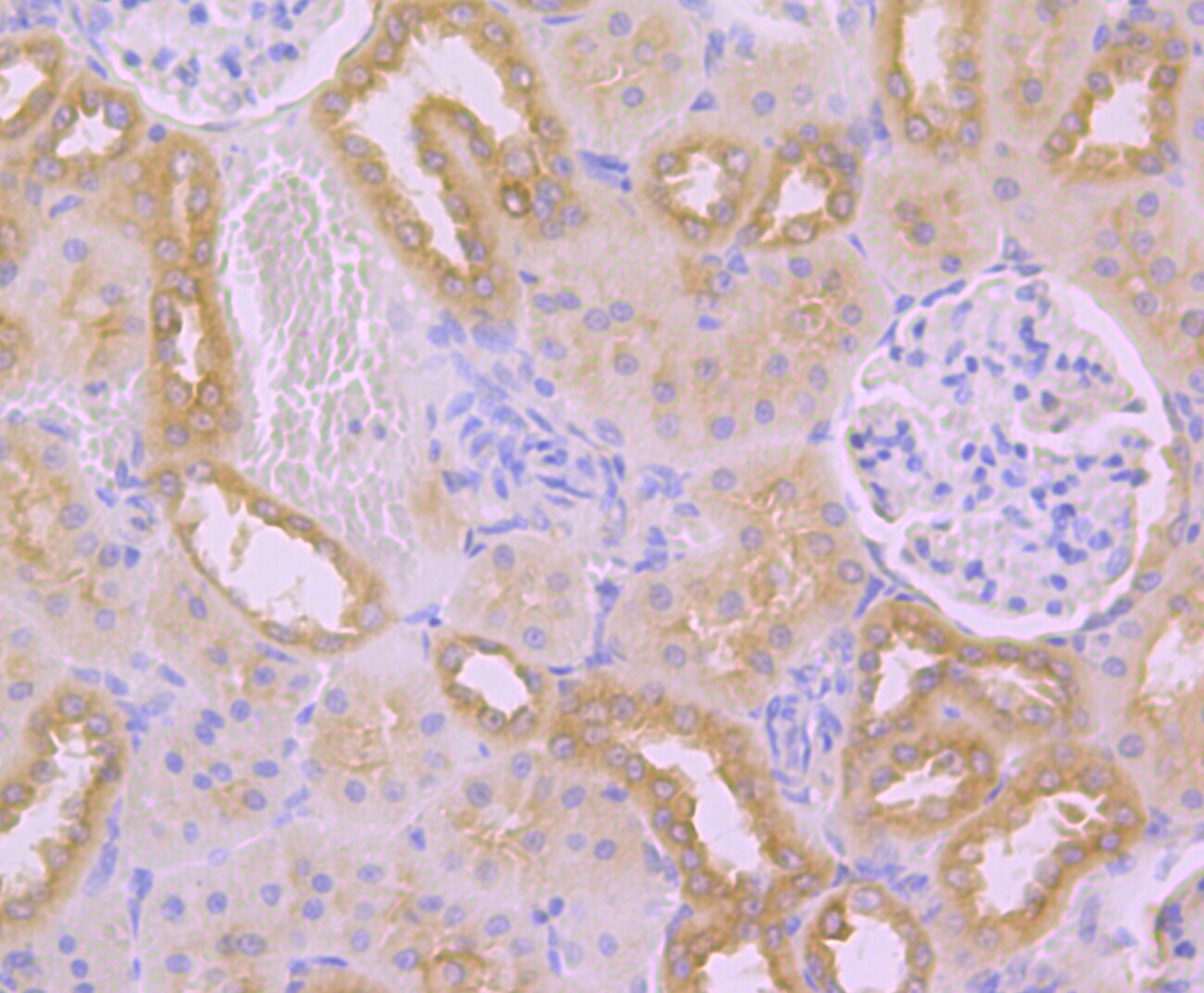
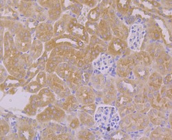
- Immunohistochemical analysis of TMEM2 of paraffin-embedded Mouse kidney tissue using a TMEM2 Polyclonal antibody (Product #PA5-85901). Counter stained with hematoxylin..

Supportive validation
- Submitted by
- Invitrogen Antibodies (provider)
- Main image
- Experimental details
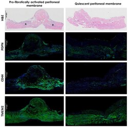
- Figure 4 HA signal transduction is involved in PA development. Peritoneal membrane from mouse PA model 3 days after induction. In H&E images, fibrotic section was apparent (*). H&E-stained samples (DiaPath, Bergamo, Italy) were imaged by a light microscope (Nikon Eclipse 50i, Tokio, Japan) with a Plan Apo 4x/0.2 Nikon JAPAN objective. Images were processed using NIS elements software (Nikon, Tokio, Japan). Nuclei of cells are stained purple, extracellular matrix and cytoplasm are stained pink. Immunofluorescent staining revealed enhanced expression pattern for HA receptor CD44 (Abcam, #ab157107, 1/500, Cambridge, GB) in profibrotically activated peritoneal membrane, while expression of typical MCs marker podoplanin (PDPN, Abcam, #ab11936, 1/100, Cambridge, GB) and hyaluronidase TMEM2 (Invitrogen, #PA5-85901, 1/100, Waltham, Massachusetts, USA) in both quiescent and pro-fibrotically activated peritoneal membranes remained homogenous. Nuclei of cells were stained using ProLong(tm) Diamond Antifade Mountant with DAPI (Invitrogen, #P36962, Waltham, MA, USA). Peritoneal membrane was imaged using a laser scanning confocal microscope (Leica TCS SP8X, Wetzlar, Germany) with the Leica HC PL APO CS2 20x/0.75 IMM (Zoom 1) objective. Images were processed using Leica LasX software and are representative of three mice. Abbreviations: PA: peritoneal adhesions, H&E: Hematoxylin and Eosin, PDPN: podoplanin, DAPI: 40,6-diamidino-2-phenylindole.
