Explore
 Validate
 Learn
Learn Western blot
Antibody data
- Antibody Data
- Antigen structure
- References [0]
- Comments [0]
- Validations
- Western blot [1]
Submit
Validation data
Reference
Comment
Report error
- Product number
- GTX17945 - Provider product page

- Provider
- GeneTex
- Proper citation
- GeneTex Cat#GTX17945, RRID:AB_422988
- Product name
- PKC mu antibody
- Antibody type
- Polyclonal
- Antigen
- Synthetic phosphopeptide derived from a region of human PKC mu that contains serine 742. The sequence is conserved in mouse. (unfortunately, the amino acid sequence is considered to be commercially sensitive)
- Host
- Rabbit
- Isotype
- IgG
- Vial size
- 50µl
- Storage
- Keep as concentrated solution. Store at 4°C short term. For extended storage aliquot and store at -20°C or below. Avoid freeze-thaw cycles.
No comments: Submit comment
Supportive validation
- Submitted by
- GeneTex (provider)
- Main image
- Experimental details
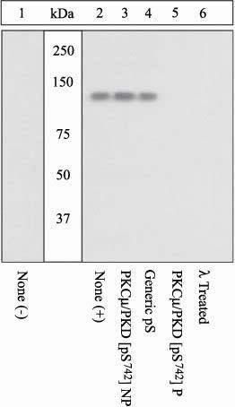
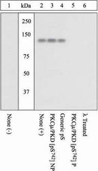
- Lysates prepared from K562 cells left unstimulated (1) or stimulated with PMA (2-6) were resolved by SDS-PAGE on a 10% polyacrylamide gel and transferred to PVDF. Membranes were either left untreated (1-5) or treated with lambda phosphatase (6), blocked with a 3% milk-TBST buffer for one hour at room temperature, and incubated with PKCµ/PKD [pS742] antibody for two hours at room temperature in a 3% milk-TBST buffer, following prior incubation with: no peptide (1, 2, 6), the non-phosphopeptide corresponding to the immunogen (3), a generic phosphoserine-containing peptide (4), or, the phosphopeptide immunogen (5). After washing, membranes were incubated with goat F(ab)2 anti-rabbit IgG HRP conjugate and bands were detected. The data show that only the peptide corresponding to PKCµ/PKD [pS742] blocks the antibody signal. The data also show that phosphatase stripping eliminates the signal, verifying that the antibody is phospho-specific
- Validation comment
- WB