Explore
 Validate
 Learn
Learn Western blot
Antibody data
- Antibody Data
- Antigen structure
- References [6]
- Comments [0]
- Validations
- Western blot [2]
- Immunocytochemistry [2]
- Other assay [5]
Submit
Validation data
Reference
Comment
Report error
- Product number
- PA1-139 - Provider product page

- Provider
- Invitrogen Antibodies
- Product name
- GADD34 Polyclonal Antibody
- Antibody type
- Polyclonal
- Antigen
- Synthetic peptide
- Description
- Western blot analysis of PA1-139 detects an ~73 kDa protein in HeLa cells. PA1-139 shows specificity to GADD34 and can be blocked by incubating with an abundance of the immunizing peptide.
- Reactivity
- Human, Mouse
- Host
- Rabbit
- Isotype
- IgG
- Vial size
- 100 μg
- Concentration
- 1 mg/mL
- Storage
- -20°C
Submitted references Inhibition of eIF2α Phosphorylation by Peste des Petits Ruminant Virus Phosphoprotein Facilitates Viral Replication.
GADD34 is a modulator of autophagy during starvation.
Raloxifene prevents stress granule dissolution, impairs translational control and promotes cell death during hypoxia in glioblastoma cells.
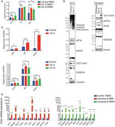
Towards Age-Related Anti-Inflammatory Therapy: Klotho Suppresses Activation of ER and Golgi Stress Response in Senescent Monocytes.
EIF1AX and RAS Mutations Cooperate to Drive Thyroid Tumorigenesis through ATF4 and c-MYC.
Heterozygous Truncating Variants in POMP Escape Nonsense-Mediated Decay and Cause a Unique Immune Dysregulatory Syndrome.
Alfred N, Qian B, Qin X, Yin X, Prajapati M, Dou Y, Li Y, Zhang Z
Frontiers in veterinary science 2021;8:645571
Frontiers in veterinary science 2021;8:645571
GADD34 is a modulator of autophagy during starvation.
Gambardella G, Staiano L, Moretti MN, De Cegli R, Fagnocchi L, Di Tullio G, Polletti S, Braccia C, Armirotti A, Zippo A, Ballabio A, De Matteis MA, di Bernardo D
Science advances 2020 Sep;6(39)
Science advances 2020 Sep;6(39)
Raloxifene prevents stress granule dissolution, impairs translational control and promotes cell death during hypoxia in glioblastoma cells.
Attwood KM, Robichaud A, Westhaver LP, Castle EL, Brandman DM, Balgi AD, Roberge M, Colp P, Croul S, Kim I, McCormick C, Corcoran JA, Weeks A
Cell death & disease 2020 Nov 17;11(11):989
Cell death & disease 2020 Nov 17;11(11):989
Towards Age-Related Anti-Inflammatory Therapy: Klotho Suppresses Activation of ER and Golgi Stress Response in Senescent Monocytes.
Mytych J, Sołek P, Będzińska A, Rusinek K, Warzybok A, Tabęcka-Łonczyńska A, Koziorowski M
Cells 2020 Jan 21;9(2)
Cells 2020 Jan 21;9(2)
EIF1AX and RAS Mutations Cooperate to Drive Thyroid Tumorigenesis through ATF4 and c-MYC.
Krishnamoorthy GP, Davidson NR, Leach SD, Zhao Z, Lowe SW, Lee G, Landa I, Nagarajah J, Saqcena M, Singh K, Wendel HG, Dogan S, Tamarapu PP, Blenis J, Ghossein RA, Knauf JA, Rätsch G, Fagin JA
Cancer discovery 2019 Feb;9(2):264-281
Cancer discovery 2019 Feb;9(2):264-281
Heterozygous Truncating Variants in POMP Escape Nonsense-Mediated Decay and Cause a Unique Immune Dysregulatory Syndrome.
Poli MC, Ebstein F, Nicholas SK, de Guzman MM, Forbes LR, Chinn IK, Mace EM, Vogel TP, Carisey AF, Benavides F, Coban-Akdemir ZH, Gibbs RA, Jhangiani SN, Muzny DM, Carvalho CMB, Schady DA, Jain M, Rosenfeld JA, Emrick L, Lewis RA, Lee B, Undiagnosed Diseases Network members, Zieba BA, Küry S, Krüger E, Lupski JR, Bostwick BL, Orange JS
American journal of human genetics 2018 Jun 7;102(6):1126-1142
American journal of human genetics 2018 Jun 7;102(6):1126-1142
No comments: Submit comment
Supportive validation
- Submitted by
- Invitrogen Antibodies (provider)
- Main image

- Experimental details
- Western blot analysis was performed on membrane enriched extracts (30 µg lysate) of HeLa (Lane 1), RAW264.7 (Lane 2), A549 (Lane 3) and NIH/3T3 (Lane 4). The blot was probed with Rabbit Anti-GADD34 Polyclonal Antibody (Product # PA1-139, 1:1000) and detected by chemiluminescence using Goat anti-Rabbit IgG (Heavy Chain) Superclonal™ Secondary Antibody, HRP conjugate (Product # A27036, 0.4 µg/mL, 1:2500 dilution). A 75 kDa band corresponding to GADD34 was observed across the cell lines tested. Known quantity of protein samples were electrophoresed using Novex® NuPAGE® 4-12 % Bis-Tris gel (Product # NP0321BOX), XCell SureLock™ Electrophoresis System (Product # EI0002) and Novex® Sharp Pre-Stained Protein Standard (Product # LC5800). Resolved proteins were then transferred onto a nitrocellulose membrane with iBlot® 2 Dry Blotting System (Product # IB21001). The membrane was probed with the relevant primary and secondary Antibody following blocking with 5 % skimmed milk. Chemiluminescent detection was performed using Pierce™ ECL Western Blotting Substrate (Product # 32106).
- Submitted by
- Invitrogen Antibodies (provider)
- Main image

- Experimental details
- Western blot analysis of GADD34 was performed by loading 10 µg of HeLa cell lysate per well onto a 4-12% Bis-Tris polyacrylamide gel. Proteins were transferred to a nitrocellulose membrane and the membrane was developed using the Fast Western Blot Kit, SuperSignal West Dura, Rabbit (Product # 35071). The membrane was probed with a GADD34 polyclonal antibody (Product # PA1-139) at dilutions ranging from 1:250-1:1000 (Lanes 2-4) for 30 minutes at room temperature on a rocking platform, washed in Fast Western Wash Buffer, and probed with Fast Western goat anti-rabbit IgG-HRP secondary antibody for at least 10 minutes. As a specificity control, PA1-139 (diluted 1:500) was pre-incubated with 5-fold molar excess of neutralizing peptide before it was used to probe the blot (Lane 5). Chemiluminescent detection was performed using SuperSignal West Dura (Product # 34075).
Supportive validation
- Submitted by
- Invitrogen Antibodies (provider)
- Main image

- Experimental details
- Immunofluorescent analysis of GADD34 (green) in HeLa cells. Formalin-fixed cells were permeabilized with 0.1% Triton X-100 in TBS for 10 minutes at room temperature and blocked with 5% normal goat serum (Product # 31873) for 15 minutes at room temperature. Cells were probed with a GADD34 polyclonal antibody (Product # PA1-139) at a dilution of 1:50 for at least 1 hour at room temperature, washed with PBS, and incubated with DyLight 488 goat anti-rabbit IgG secondary antibody (Product # 35552) at a dilution of 1:500 for 30 minutes at room temperature. F-actin (red) was stained with DyLight 650 Phalloidin (Product # 21838) and nuclei (blue) were stained with Hoechst 33342 dye (Product # 62249). Images were taken on a Thermo Scientific ArrayScan or ToxInsight Instrument at 20X magnification.

- Submitted by
- Invitrogen Antibodies (provider)
- Main image
- Experimental details
- Immunofluorescent analysis of GADD34 (green) in HeLa cells. Formalin-fixed cells were permeabilized with 0.1% Triton X-100 in TBS for 10 minutes at room temperature and blocked with 5% normal goat serum (Product # 31873) for 15 minutes at room temperature. Cells were probed with a GADD34 polyclonal antibody (Product # PA1-139) at a dilution of 1:50 for at least 1 hour at room temperature, washed with PBS, and incubated with DyLight 488 goat anti-rabbit IgG secondary antibody (Product # 35552) at a dilution of 1:500 for 30 minutes at room temperature. F-actin (red) was stained with DyLight 650 Phalloidin (Product # 21838) and nuclei (blue) were stained with Hoechst 33342 dye (Product # 62249). Images were taken on a Thermo Scientific ArrayScan or ToxInsight Instrument at 20X magnification.

Supportive validation
- Submitted by
- Invitrogen Antibodies (provider)
- Main image
- Experimental details
- NULL
- Submitted by
- Invitrogen Antibodies (provider)
- Main image
- Experimental details
- Fig. 5 Raloxifene delays resumption of protein translation post-hypoxia in correlation with increased eIF2alpha phosphorylation and decreased mTOR signaling. A and B Puromycin incorporation assay. U251 cells were treated with 40 muM raloxifene or DMSO vehicle control for 1 h prior to 2 h hypoxic A or non-hypoxic B incubation. At various times post-hypoxic or normoxic incubation cells were treated with 10 mug/mL puromycin for 10 min and puromycin incorporation into nascent proteins was detected by anti-puromycin blot. Blots were normalized to total lane protein, and represented as ratios with DMSO time 0 normalized to 1 (bottom quantification panels). Data is presented as the mean of triplicates +- SEM, unpaired t -test * p < 0.05; ** p < 0.01. C and D U251 cells were treated as in A but without addition of puromycin. Cell lysates were probed for eIF2alpha, phospho-eIF2alpha, GADD34, rpS6, and phospho-rpS6 at various times post-hypoxic C or normoxic D incubation. All blots were normalized to total lane protein with DMSO time 0 set to 1 (bottom quantification panels). The level of eIF2alpha phosphorylation is presented as the ratio of p-eIF2alpha to total eIF2alpha (bottom quantification panels). The level of rpS6 phosphorylation is similarly presented. Data is presented as the mean of triplicates +- SEM, unpaired t -test, and false discovery rate of 1%. * p < 0.05; ** p < 0.01.
- Submitted by
- Invitrogen Antibodies (provider)
- Main image
- Experimental details
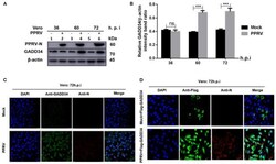
- Figure 2 PPRV infection modulates GADD34 protein expression in Vero cells. Vero cells were infected with PPRV or mock-infected at indicated times. (A) The effect of PPRV on GADD34 protein expression was analyzed by Western blot. (B) Densitometry quantification of GADD34 was normalized to beta-actin and fold change was measured within each time point compared with mock-infected by ImageJ analysis. Statistical analysis was performed using GraphPad Prism 8 software. Shown are representative immunoblots and the error bars represent the mean +- SD from three independent experiments. Asterisks represent the statistically significant difference between mock and infected cells (*** P < 0.001 and ns, not significant). (C) Vero cells were infected with PPRV or mock-infected for 72 h. (D) Vero cells were transfected with Flag-GADD34 (12 h) before infection with PPRV or mock infection for a further 72 h. Cells were double-stained with mouse monoclonal antibodies against PPRV N protein (1:50) and Rabbit anti-GADD34 antibodies (PA1-139) 1:100 in (C) or Rabbit anti-Flag (ab1162) 1:200 in (D) as primary antibodies overnight. Goat anti-rabbit IgG (H&L) Alexa Fluor 488 (ab150077) Green1:500 and Goat anti-mouse IgG H&L (Alexa Fluor 647) (ab150115) Red 1:1000 were used as secondary antibodies for 1 h at room temperature. DAPI was used to stain the cell nuclei (blue).
- Submitted by
- Invitrogen Antibodies (provider)
- Main image

- Experimental details
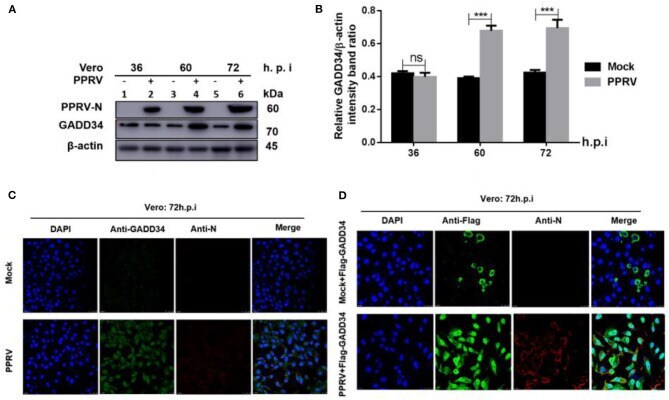
- Figure 3 PPRV infection represses ER stress-induced eIF2alpha phosphorylation in Vero cells. Vero cells were infected with PPRV or mock-infected. (A,C) PPRV infection represses eIF2alpha phosphorylation, induced by DTT and TG, respectively. PPRV-infected or mock-infected cells were treated with DTT (2 mM) or TG (1 muM) for 2 h (for DTT) or 12 h (for TG), respectively, to induce ER stress. The effect of PPRV on p-eIF2alpha and GADD34 expression in DTT- or TG-treated groups were analyzed by Western blot. (B,D) Densitometry quantification of p-eIF2alpha and GADD34 was normalized to beta-actin. Fold change was measured and compared with mock-infected treated or untreated by ImageJ analysis. Statistical analysis was performed using GraphPad Prism 8.0 software. Shown are representative immunoblots with the error bars representing the mean +- SD from three independent experiments. Asterisks represent statistically significant differences between treated and untreated groups (* P < 0.05; ** P < 0.01). # In the graphic (D) , plot 5 represents one set of untreated samples (lines 5 to 7) and plot 6 represents one set of TG-treated samples (lines 8 to 10).
- Submitted by
- Invitrogen Antibodies (provider)
- Main image
- Experimental details
- Figure 4 Inhibition of eIF2alpha dephosphorylation is beneficial to PPRV replication. Vero cells were infected with PPRV or mock-infected. (A,C) After 2-h adsorption, infection media was replaced by fresh media treated with 5, 10, and 25 muM of Salubrinal in (A) or ISRIB (200 nM) in (C) and incubated up to 60 h.p.i. for Salubrinal and 48 h.p.i. for ISRIB. Treatment with DMSO alone was used as control. The effect of PPRV on p-eIF2alpha in treated or untreated groups were analyzed by Western blot. (B,D) Densitometry quantification of p-eIF2alpha, GADD34, and PPRV-N was normalized to beta-actin and fold change between treated and untreated was determined by ImageJ analysis. Statistical analysis was performed using GraphPad Prism 8.0 software. On the graphs, shown are representative immunoblots with the error bars representing the mean +- SD from three independent experiments. (E,F) RT-qPCR was used to quantify the mRNA levels of PPRV in samples treated or untreated with Salubrinal (E) or ISRIB (F) . Viral RNA of the positive control (PC) or vehicle treated with DMSO were compared to that of Salubrinal or ISRIB treated groups. The mean +- standard deviation (SD) was obtained in triplicate ( n = 3). Statistical analysis was performed using GraphPad Prism 8.0 software using multiple t -tests. Asterisks represent statistically significant differences between treated and untreated groups (* P < 0.05; ** P < 0.01; *** P < 0.001; **** P < 0.0001; ns, not significant).