 Explore
 Validate
 Learn
Learn Western blot
 Immunocytochemistry
Antibody data
- Antibody Data
- Antigen structure
- References [5]
- Comments [0]
- Validations
- Western blot [4]
- Immunocytochemistry [1]
- Immunohistochemistry [1]
Submit
Validation data
Reference
Comment
Report error
- Product number
- GTX101803 - Provider product page

- Provider
- GeneTex
- Proper citation
- GeneTex Cat#GTX101803, RRID:AB_1950200
- Product name
- ENO1 antibody [N3C3]
- Antibody type
- Polyclonal
- Reactivity
- Human, Mouse
- Host
- Rabbit
Submitted references Proteomic investigating the cooperative lethal effect of EGFR and MDM2 inhibitors on ovarian carcinoma.
Identification of Differentiation-Related Proteins in Gastric Adenocarcinoma Tissues by Proteomics.
Integrating transcriptomics and proteomics to show that tanshinone IIA suppresses cell growth by blocking glucose metabolism in gastric cancer cells.
Quantitative proteomic analysis of human lung tumor xenografts treated with the ectopic ATP synthase inhibitor citreoviridin.
Surface α-enolase promotes extracellular matrix degradation and tumor metastasis and represents a new therapeutic target.
Chang SJ, Liao EC, Yeo HY, Kuo WH, Chen HY, Tsai YT, Wei YS, Chen YJ, Wang YS, Li JM, Shih CC, Chan CH, Lai ZY, Chou HC, Chuang YJ, Chan HL
Archives of biochemistry and biophysics 2018 Jun 1;647:10-32
Archives of biochemistry and biophysics 2018 Jun 1;647:10-32
Identification of Differentiation-Related Proteins in Gastric Adenocarcinoma Tissues by Proteomics.
Zhou X, Yao K, Zhang L, Zhang Y, Han Y, Liu HL, Liu XW, Su G, Yuan WZ, Wei XD, Guan QL, Zhu BD
Technology in cancer research & treatment 2016 Oct;15(5):697-706
Technology in cancer research & treatment 2016 Oct;15(5):697-706
Integrating transcriptomics and proteomics to show that tanshinone IIA suppresses cell growth by blocking glucose metabolism in gastric cancer cells.
Lin LL, Hsia CR, Hsu CL, Huang HC, Juan HF
BMC genomics 2015 Feb 5;16:41
BMC genomics 2015 Feb 5;16:41
Quantitative proteomic analysis of human lung tumor xenografts treated with the ectopic ATP synthase inhibitor citreoviridin.
Wu YH, Hu CW, Chien CW, Chen YJ, Huang HC, Juan HF
PloS one 2013;8(8):e70642
PloS one 2013;8(8):e70642
Surface α-enolase promotes extracellular matrix degradation and tumor metastasis and represents a new therapeutic target.
Hsiao KC, Shih NY, Fang HL, Huang TS, Kuo CC, Chu PY, Hung YM, Chou SW, Yang YY, Chang GC, Liu KJ
PloS one 2013;8(7):e69354
PloS one 2013;8(7):e69354
No comments: Submit comment
Enhanced validation
Supportive validation
- Submitted by
- GeneTex (provider)
- Enhanced method
- Genetic validation
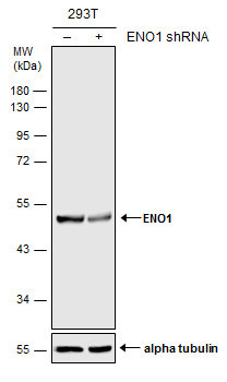
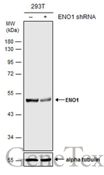
- Main image
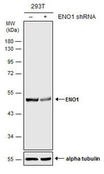
- Experimental details
- Non-transfected (¡V) and transfected (+) 293T whole cell extracts (30 ?g) were separated by 10% SDS-PAGE, and the membrane was blotted with ENO1 antibody [N3C3] (GTX101803) diluted at 1:20000. The HRP-conjugated anti-rabbit IgG antibody (GTX213110-01) was used to detect the primary antibody.
Supportive validation
- Submitted by
- GeneTex (provider)
- Main image
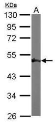
- Experimental details
- Sample (50 ?g of whole cell lysate) A: Mouse brain 10% SDS PAGE GTX101803 diluted at 1:1000 The HRP-conjugated anti-rabbit IgG antibody (GTX213110-01) was used to detect the primary antibody.

- Submitted by
- GeneTex (provider)
- Main image
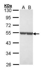
- Experimental details
- Sample (30 ?g of whole cell lysate) A: A431 (GTX27909) B: H1299 10% SDS PAGE GTX101803 diluted at 1:3000The HRP-conjugated anti-rabbit IgG antibody (GTX213110-01) was used to detect the primary antibody.

- Submitted by
- GeneTex (provider)
- Main image
- Experimental details
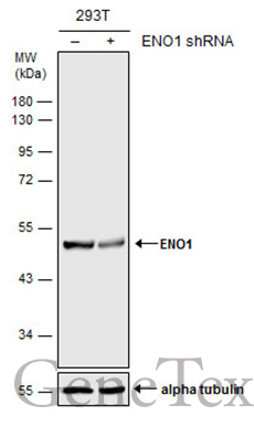
- Non-transfected (¡V) and transfected (+) 293T whole cell extracts (30 ?g) were separated by 10% SDS-PAGE, and the membrane was blotted with ENO1 antibody [N3C3] (GTX101803) diluted at 1:20000. The HRP-conjugated anti-rabbit IgG antibody (GTX213110-01) was used to detect the primary antibody.
Supportive validation
- Submitted by
- GeneTex (provider)
- Main image
- Experimental details
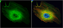
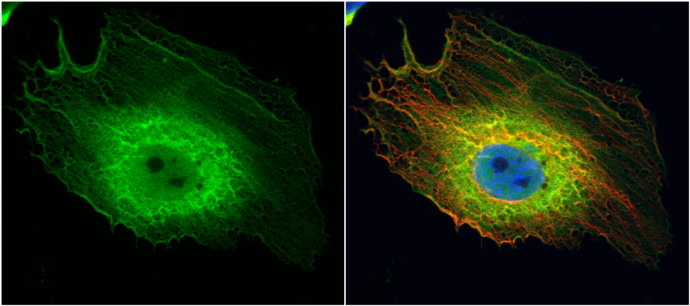
- ENO1 antibody [N3C3] detects ENO1 protein at cytoplasm by immunofluorescent analysis.Sample: HeLa cells were fixed in 4% paraformaldehyde at RT for 15 min.Green: ENO1 protein stained by ENO1 antibody [N3C3] (GTX101803) diluted at 1:200.Red: alpha Tubulin, a cytoskeleton marker, stained by alpha Tubulin antibody [B-5-1-2] (GTX11304) diluted at 1:10000.Blue: Hoechst 33342 staining.
Supportive validation
- Submitted by
- GeneTex (provider)
- Main image
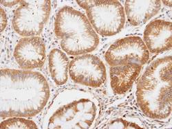
- Experimental details
- Immunohistochemical analysis of paraffin-embedded human gastric tissue, using ENO1(GTX101803) antibody at 1:100 dilution.
