Explore
 Validate
 Learn
Learn Western blot
 Immunocytochemistry
Antibody data
- Antibody Data
- Antigen structure
- References [1]
- Comments [0]
- Validations
- Immunocytochemistry [2]
- Flow cytometry [1]
- Other assay [1]
Submit
Validation data
Reference
Comment
Report error
- Product number
- PA5-13459 - Provider product page

- Provider
- Invitrogen Antibodies
- Product name
- ENO1 Polyclonal Antibody
- Antibody type
- Polyclonal
- Antigen
- Synthetic peptide
- Description
- This antibody is predicted to react with bovine, chicken, non-human primate, rat and Xenopus based on sequence homology.
- Reactivity
- Human, Mouse
- Host
- Rabbit
- Isotype
- IgG
- Vial size
- 400 µL
- Concentration
- 2 mg/mL
- Storage
- Store at 4°C short term. For long term storage, store at -20°C, avoiding freeze/thaw cycles.
Submitted references Alpha-Enolase (ENO1) Correlates with Invasiveness of Cutaneous Melanoma-An In Vitro and a Clinical Study.
Hippner M, Majkowski M, Biecek P, Szkudlarek T, Simiczyjew A, Pieniazek M, Nowak D, Miazek A, Donizy P
Diagnostics (Basel, Switzerland) 2022 Jan 20;12(2)
Diagnostics (Basel, Switzerland) 2022 Jan 20;12(2)
No comments: Submit comment
Supportive validation
- Submitted by
- Invitrogen Antibodies (provider)
- Main image
- Experimental details
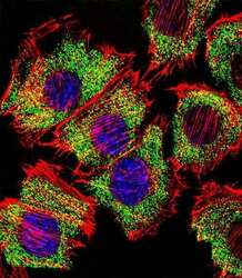
- Immunofluorescent analysis of C2C12 cells stained with an ENO1 polyclonal antibody (Product # PA5-13459). C2C12 cells were fixed with 4% PFA (20 min), permeabilized with Triton X-100 (0.1%, 10 min), then incubated with an ENO1 polyclonal antibody (Product # PA5-13459) (1:25, 1 hr at 37°C). Primary antibody was detected with fluor-conjugated donkey anti-rabbit secondary antibody (green) at 1:400 dilution for 50 min at 37°C). Actin filaments have been labeled with dye-conjugated phalloidin (red). Nuclei were counterstained with DAPI (blue) (10 µg/mL, 10 min).

- Submitted by
- Invitrogen Antibodies (provider)
- Main image
- Experimental details
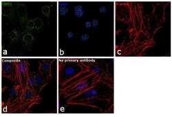
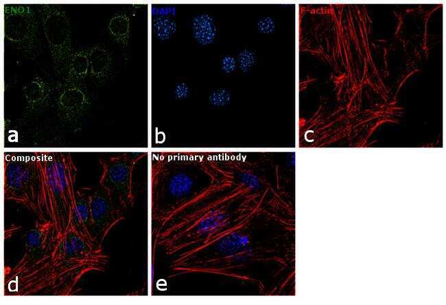
- Immunofluorescence analysis of ENO1 was performed using 70% confluent log phase C2C12 cells. The cells were fixed with 4% paraformaldehyde for 10 minutes, permeabilized with 0.1% Triton™ X-100 for 15 minutes, and blocked with 1% BSA for 1 hour at room temperature. The cells were labeled with ENO1 Polyclonal Antibody (Product # PA5-13459) at 1:100 dilution in 0.1% BSA, incubated at 4 degree Celsius overnight and then labeled with Goat anti-Rabbit IgG (H+L) Superclonal™ Secondary Antibody, Alexa Fluor® 488 conjugate (Product # A27034) at a dilution of 1:2000 for 45 minutes at room temperature (Panel a: green). Nuclei (Panel b: blue) were stained with SlowFade® Gold Antifade Mountant with DAPI (Product # S36938). F-actin (Panel c: red) was stained with Rhodamine Phalloidin (Product # R415, 1:300). Panel d represents the merged image showing cytoplasmic localization. Panel e represents control cells with no primary antibody to assess background. The images were captured at 60X magnification.
Supportive validation
- Submitted by
- Invitrogen Antibodies (provider)
- Main image
- Experimental details
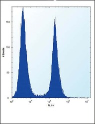
- Flow cytometry analysis of HeLa cells using an ENO1 polyclonal antibody (Product # PA5-13459) (right) compared to a negative control cell (left) at a dilution of 1:10-50, followed by a FITC-conjugated donkey anti-rabbit antibody

Supportive validation
- Submitted by
- Invitrogen Antibodies (provider)
- Main image
- Experimental details
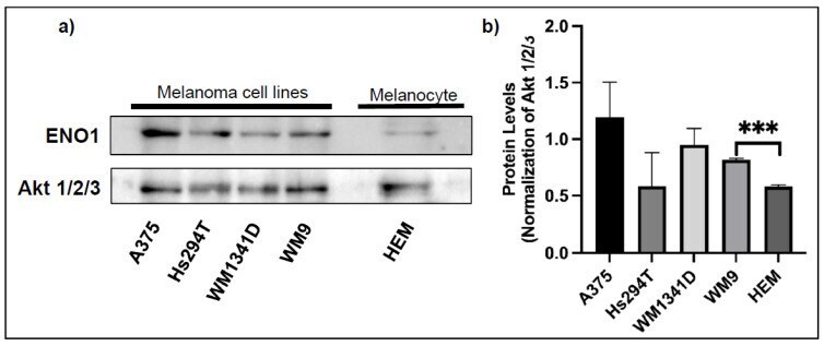
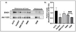
- Figure 1 The expression level of ENO1 in the cell lysates from primary melanocytes and melanoma cell lines. Representative Western blots showing ENO1 and Akt 1/2/3 expression (for normalization) in protein lysates obtained from the primary human melanocytes (HEM) and indicated melanoma cell lines ( a ). Densitometric ENO1/Akt ratios are shown as mean values ( n = 3 except for HEM, n = 2) +- standard error of the mean (SEM) ( b ). The significance level was set at p = 0.001-0.0001 (***).