Explore
 Validate
 Learn
Learn Western blot
Antibody data
- Antibody Data
- Antigen structure
- References [1]
- Comments [0]
- Validations
- Western blot [4]
- Immunocytochemistry [1]
- Immunohistochemistry [1]
- Other assay [2]
Submit
Validation data
Reference
Comment
Report error
- Product number
- PA5-29660 - Provider product page

- Provider
- Invitrogen Antibodies
- Product name
- ENO1 Polyclonal Antibody
- Antibody type
- Polyclonal
- Antigen
- Recombinant protein fragment
- Description
- Recommended positive controls: 293T.
- Concentration
- 1 mg/mL
Submitted references Quantitative Proteomic Approach Reveals Altered Metabolic Pathways in Response to the Inhibition of Lysine Deacetylases in A549 Cells under Normoxia and Hypoxia.
Martín-Bernabé A, Tarragó-Celada J, Cunin V, Michelland S, Cortés R, Poignant J, Boyault C, Rachidi W, Bourgoin-Voillard S, Cascante M, Seve M
International journal of molecular sciences 2021 Mar 25;22(7)
International journal of molecular sciences 2021 Mar 25;22(7)
No comments: Submit comment
Supportive validation
- Submitted by
- Invitrogen Antibodies (provider)
- Main image
- Experimental details
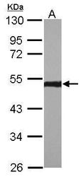
- Western blot analysis of ENO1 using 30 µg of A431 lysate. Samples were loaded onto a 10% SDS-PAGE gel and probed with an ENO1 polyclonal antibody (Product # PA5-29660) at a dilution of 1:1000.

- Submitted by
- Invitrogen Antibodies (provider)
- Main image
- Experimental details
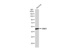
- Western Blot using ENO1 Polyclonal Antibody (Product # PA5-29660). Rat tissue extract (50 µg) was separated by 10% SDS-PAGE, and the membrane was blotted with ENO1 Polyclonal Antibody (Product # PA5-29660) diluted at 1:1,000. The HRP-conjugated anti-rabbit IgG antibody was used to detect the primary antibody.

- Submitted by
- Invitrogen Antibodies (provider)
- Main image

- Experimental details
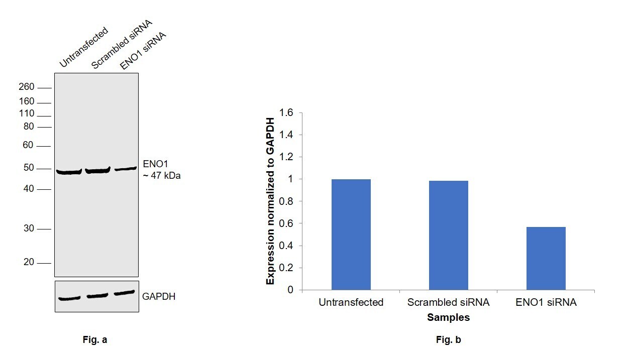
- Knockdown of Alpha-enolase (ENO1) was achieved by transfecting U-87 MG cells with ENO1 specific siRNAs (Silencer® select Product # s4680). Western blot analysis (Fig. a) was performed using whole cell extracts from the ENO1 knockdown cells (lane 3), non-specific scrambled siRNA transfected cells (lane 2) and untransfected cells (lane 1). The blot was probed using ENO1 Polyclonal Antibody (Product # PA5-29660, 1:1000 dilution) and Goat anti-Rabbit IgG (H+L) Superclonal™ Recombinant Secondary Antibody, HRP (Product # A27036, 0.25 µg/mL, 1:4000 dilution). Densitometric analysis of this western blot is shown in histogram (Fig. b). Decrease in signal upon siRNA mediated knock down confirms that antibody is specific to ENO1
- Submitted by
- Invitrogen Antibodies (provider)
- Main image
- Experimental details
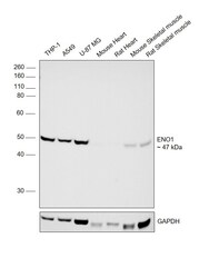
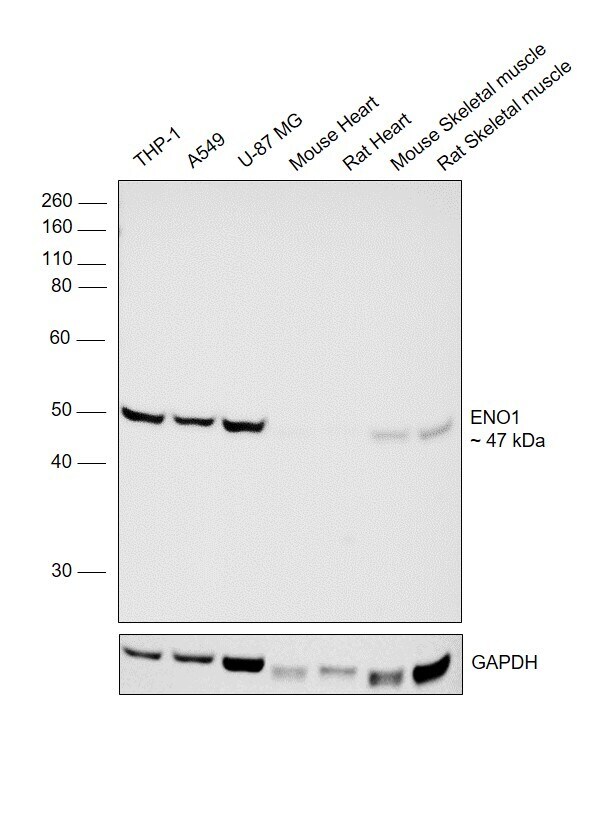
- Western blot was performed using anti-ENO1 Polyclonal Antibody (Product # PA5-29660) on whole cell extracts (30 µg lysate) of THP-1 (Lane1), A549 (Lane 2), U-87 MG (Lane 3), Mouse Heart (Lane 4), Rat Heart (Lane 5), Mouse Skeletal muscle (Lane 6) and Rat Skeletal muscle (Lane 7) and 47 kDa band corresponding to ENO1 protein was observed except in Mouse and Rat Heart. Resolved proteins were then transferred onto a nitrocellulose membrane (Product # IB23001) by iBlot® 2 Dry Blotting System (Product # IB21001). The blot was probed with the primary antibody (1:1000 dilution) and detected by chemiluminescence with Goat anti-Rabbit IgG (H+L) Superclonal™ Recombinant Secondary Antibody, HRP (Product # A27036, 0.25 µg/mL, 1:4000 dilution) using the iBright FL 1000 (Product # A32752). Chemiluminescent detection was performed using Novex® ECL Chemiluminescent Substrate Reagent Kit (Product # WP20005).
Supportive validation
- Submitted by
- Invitrogen Antibodies (provider)
- Main image
- Experimental details
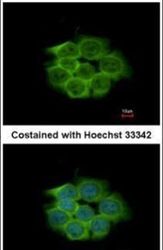
- Immunofluorescent analysis of ENO1 in methanol-fixed HCT116 cells using an ENO1 polyclonal antibody (Product # PA5-29660) at a 1:500 dilution.

Supportive validation
- Submitted by
- Invitrogen Antibodies (provider)
- Main image
- Experimental details
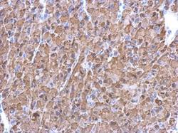
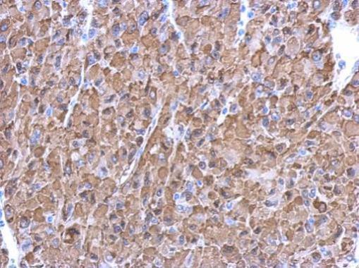
- Immunohistochemical analysis of paraffin-embedded U87 xenograft, using ENO1 (Product # PA5-29660) antibody at 1:500 dilution. Antigen Retrieval: EDTA based buffer, pH 8.0, 15 min.
Supportive validation
- Submitted by
- Invitrogen Antibodies (provider)
- Main image
- Experimental details
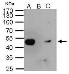
- ENO1antibody immunoprecipitates ENO1protein in IP experiments. IP Sample: 1,000 µg HeLa whole cell lysate/extract A. 40 µg HeLa whole cell lysate/extract B. Control with 2.5 µg of preimmune rabbit IgG C. Immunoprecipitation of ENO1protein by 2.5 µg of ENO1 Polyclonal Antibody (Product # PA5-29660) 10% SDS-PAGE The immunoprecipitated ENO1 protein was detected by ENO1 Polyclonal Antibody (Product # PA5-29660) diluted at 1:1,000.

- Submitted by
- Invitrogen Antibodies (provider)
- Main image
- Experimental details
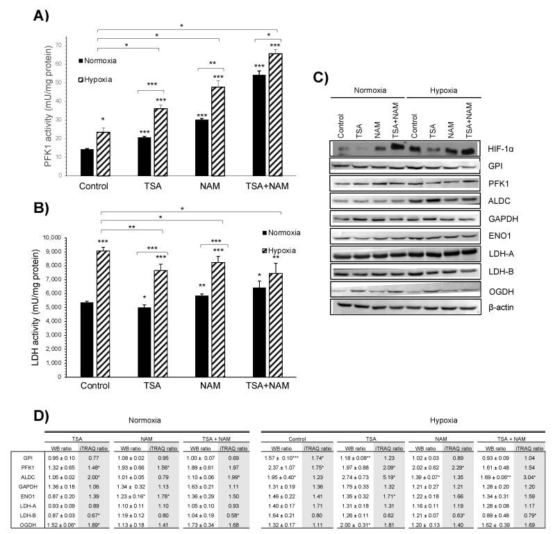
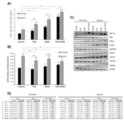
- Figure 4 Effect of KDAC inhibition on enzyme activities in A549 cells under normoxia and hypoxia. ( A , B ) The ATP- dependent 6-phosphofructokinase (PFK1) ( A ) and lactate dehydrogenase (LDH) ( B ) enzymatic activities were measured after 24 h of incubation, and activities were normalized to intracellular protein content in each condition. A549 cells were treated with 1 muM of TSA, 20 mM of NAM, and both 1 muM TSA and 20 mM NAM for 24 h of incubation under normoxia and hypoxia. Cells incubated in medium without KDACIs served as control. Bars represent the means +- standard error of the mean of three independent experiments. The asterisks above bars indicate statistically significant differences compared to normoxic control cells. The asterisks above curly brackets indicate statistically significant differences between hypoxic and normoxic treatments and between hypoxic treatments and hypoxic control cells. Statistical significance was assessed by a two-tailed Student''s t -test. *, p