Explore
 Validate
 Learn
LearnMA5-31959
antibody from Invitrogen Antibodies
Targeting: LRP1
A2MR, APOER, APR, CD91, IGFBP-3R, IGFBP3R1, LRP, LRP1A
 Western blot
Antibody data
- Antibody Data
- Antigen structure
- References [2]
- Comments [0]
- Validations
- Western blot [3]
- Immunocytochemistry [6]
- Immunoprecipitation [1]
- Immunohistochemistry [8]
- Flow cytometry [1]
- Other assay [1]
Submit
Validation data
Reference
Comment
Report error
- Product number
- MA5-31959 - Provider product page

- Provider
- Invitrogen Antibodies
- Product name
- LRP1 Recombinant Rabbit Monoclonal Antibody (SA0290)
- Antibody type
- Monoclonal
- Antigen
- Synthetic peptide
- Description
- Recombinant rabbit monoclonal antibodies are produced using in vitro expression systems. The expression systems are developed by cloning in the specific antibody DNA sequences from immunoreactive rabbits. Then, individual clones are screened to select the best candidates for production. The advantages of using recombinant rabbit monoclonal antibodies include: better specificity and sensitivity, lot-to-lot consistency, animal origin-free formulations, and broader immunoreactivity to diverse targets due to larger rabbit immune repertoire.
- Reactivity
- Human, Mouse, Rat
- Host
- Rabbit
- Isotype
- IgG
- Antibody clone number
- SA0290
- Vial size
- 100 μL
- Concentration
- 1 mg/mL
- Storage
- Store at 4°C short term. For long term storage, store at -20°C, avoiding freeze/thaw cycles.
Submitted references LDL receptor-related protein 1 (LRP1), a novel target for opening the blood-labyrinth barrier (BLB).
CREBH normalizes dyslipidemia and halts atherosclerosis in diabetes by decreasing circulating remnant lipoproteins.
Shi X, Wang Z, Ren W, Chen L, Xu C, Li M, Fan S, Xu Y, Chen M, Zheng F, Zhang W, Zhou X, Zhang Y, Qiu S, Wu L, Zhou P, Lv X, Cui T, Qiao Y, Zhao H, Guo W, Chen W, Li S, Zhong W, Lin J, Yang S
Signal transduction and targeted therapy 2022 Jun 10;7(1):175
Signal transduction and targeted therapy 2022 Jun 10;7(1):175
CREBH normalizes dyslipidemia and halts atherosclerosis in diabetes by decreasing circulating remnant lipoproteins.
Shimizu-Albergine M, Basu D, Kanter JE, Kramer F, Kothari V, Barnhart S, Thornock C, Mullick AE, Clouet-Foraison N, Vaisar T, Heinecke JW, Hegele RA, Goldberg IJ, Bornfeldt KE
The Journal of clinical investigation 2021 Nov 15;131(22)
The Journal of clinical investigation 2021 Nov 15;131(22)
No comments: Submit comment
Supportive validation
- Submitted by
- Invitrogen Antibodies (provider)
- Main image
- Experimental details
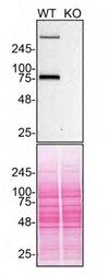
- Western blot of LRP1 was performed by loading 30 µg of HAP1 WT (lane 1) and HAP1 LRP1 KO (lane 2) cell lysates in RIPA buffer (Product # 89901) onto a 4-20% gradient polyacrylamide gel (Product # WXP42012BOX). Proteins on the blot were visualized with Ponceau staining (below immunoblot) (Product # BP10310). Proteins were transferred to nitrocellulose membrane and blocked in 5% milk for 1 hr. LRP1 was detected at approximately 85 kDa using LRP1 Recombinant Rabbit Monoclonal Antibody (SA0290) (Product # MA5-31959) at a dilution of 1:2,000 in 5% BSA in TBST overnight at 4 degree celsius. The blot was probed with Goat anti-Rabbit IgG (H+L) Secondary Antibody, HRP (Product # 65-6120) diluted to 0.2 µg/mL in TBST with 5% milk for 1 hr at room temperature. Chemiluminescent detection was performed using ECL Western Blotting Substrate and iBright™ CL1500 Imaging System (Product # A44240). Data courtesy of YCharOS Inc., an open science company with the mission of characterizing commercially available antibodies using knockout validation.

- Submitted by
- Invitrogen Antibodies (provider)
- Main image
- Experimental details
- Immunohistochemical analysis of TCF7L2 in Human breast cancer tissue using a TCF7L2 Polyclonal antibody (Product #PA5-85993). Counter stained with hematoxylin..

- Submitted by
- Invitrogen Antibodies (provider)
- Main image
- Experimental details
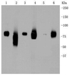
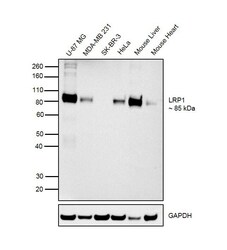
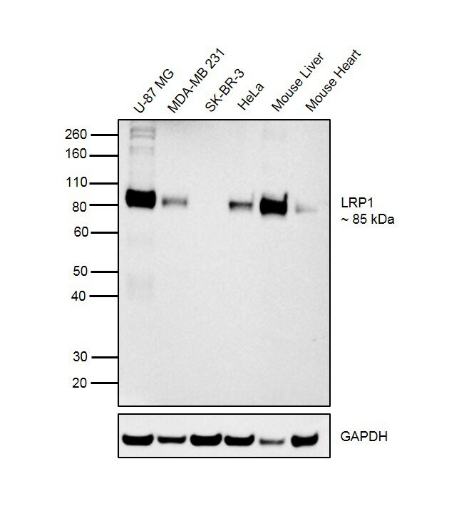
- Western blot was performed using Anti-LRP1 Recombinant Rabbit Monoclonal Antibody (Product # MA5-31959) and a 85 kDa band corresponding to LRP1 was observed in cell lines and tissues tested except SK-BR-3 cell line. Whole cell extracts (30 µg lysate) of U-87 MG (Lane 1), MDA-MB-231 (Lane 2), SK-BR-3 (Lane 3), HeLa (Lane 4), Mouse Liver (Lane 5) and Mouse Heart (Lane 6) were electrophoresed using Novex® NuPAGE® 4-12% Bis-Tris Protein Gel (Product # NP0322BOX). Resolved proteins were then transferred onto a nitrocellulose membrane (Product # IB23001) by iBlot® 2 Dry Blotting System (Product # IB21001). The blot was probed with the primary antibody (1:2500 dilution) and detected by Goat anti-Rabbit IgG (Heavy Chain) Superclonal™ Recombinant Secondary Antibody, HRP (Product # A27036, 1:4000 dilution) using the iBright FL 1000 (Product # A32752). Chemiluminescent detection was performed using Novex® ECL Chemiluminescent Substrate Reagent Kit (Product # WP20005).
Supportive validation
- Submitted by
- Invitrogen Antibodies (provider)
- Main image
- Experimental details
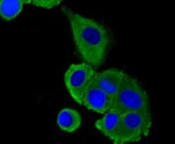
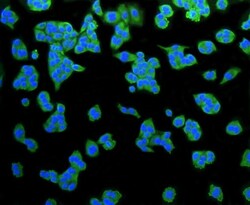
- Immunocytochemical analysis of LRP1 in MCF-7 cells using a LRP1 Monoclonal antibody (Product # MA5-31959) as seen in green. The nuclear counter stain is DAPI (blue). Cells were fixed in paraformaldehyde, permeabilised with 0.25% Triton X100/PBS.

- Submitted by
- Invitrogen Antibodies (provider)
- Main image

- Experimental details
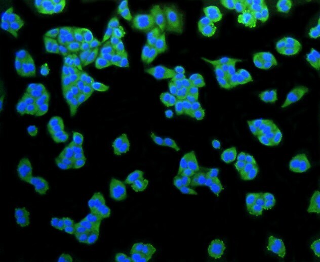
- Immunocytochemical analysis of LRP1 in HUVEC cells using a LRP1 Monoclonal antibody (Product # MA5-31959) as seen in green. Cells were fixed in paraformaldehyde, permeabilised with 0.25% Triton X100/PBS.

- Submitted by
- Invitrogen Antibodies (provider)
- Main image

- Experimental details
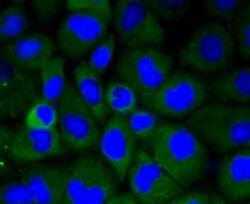
- Immunocytochemical analysis of LRP1 in Hela cells using a LRP1 Monoclonal antibody (Product # MA5-31959) as seen in green. The nuclear counter stain is DAPI (blue). Cells were fixed in paraformaldehyde, permeabilised with 0.25% Triton X100/PBS.

- Submitted by
- Invitrogen Antibodies (provider)
- Main image
- Experimental details
- Immunocytochemical analysis of LRP1 in HUVEC cells using a LRP1 Monoclonal antibody (Product # MA5-31959) as seen in green. Cells were fixed in paraformaldehyde, permeabilised with 0.25% Triton X100/PBS.
- Submitted by
- Invitrogen Antibodies (provider)
- Main image
- Experimental details
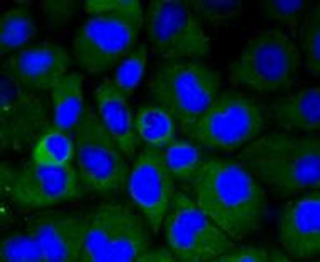
- Immunocytochemical analysis of LRP1 in Hela cells using a LRP1 Monoclonal antibody (Product # MA5-31959) as seen in green. The nuclear counter stain is DAPI (blue). Cells were fixed in paraformaldehyde, permeabilised with 0.25% Triton X100/PBS.
- Submitted by
- Invitrogen Antibodies (provider)
- Main image
- Experimental details
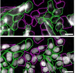
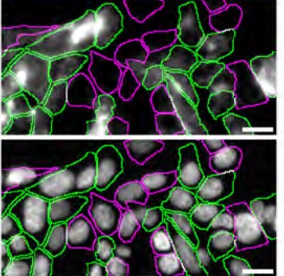
- Immunofluorescence of LRP1 was performed using HAP1 WT and HAP1 LRP1 KO cells that were labeled with a green or a far-red fluorescent dye, respectively. WT and KO cells were mixed and plated to a 1:1 ratio on glass coverslips as a mosaic and incubated for 24 hrs. Cells were fixed in 4% PFA (in PBS) for 15 min at RT; cells were permeabilized with 0.1% Triton X-100 for 10 min at RT and blocked with PBS containing 5% BSA, 5% goat serum, and 0.01% Triton X-100 for 30 min. Cells were stained with LRP1 Recombinant Rabbit Monoclonal Antibody (SA0290) (Product # MA5-31959) at a 1:200 dilution overnight at 4 degree celcius. Secondary antibody incubation was performed using 1 µg/mL of Goat anti-Rabbit IgG (H+L) Highly Cross-Adsorbed Secondary Antibody, Alexa Fluor™ 555 (Product # A-21429) together with DAPI for 1 hr at RT. Imaging was performed with a 20X water immersion objective. Representative images where WT and KO cells are outlined with a green (WT) or magenta (KO) line, respectively, are shown. The top and bottom panels show antibody and DAPI stainings, respectively. Scale bar = 10 µm. Data courtesy of YCharOS Inc., an open science company with the mission of characterizing commercially available antibodies using knockout validation.
Supportive validation
- Submitted by
- Invitrogen Antibodies (provider)
- Main image
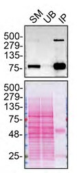
- Experimental details
- Immunoprecipitation of LRP1 was performed on HAP1 WT cell lysate. Antibody-bead conjugate was prepared by adding 2 µg of LRP1 Recombinant Rabbit Monoclonal Antibody (SA0290) (Product # MA5-31959) and 30 µL of Dynabeads™ Protein A (Product # 10002D) to 500 µl of Pierce™ IP Lysis Buffer (Product # 87788). The mixture was rocked for ~1 hour at 4 degree celcius followed by two washes to remove unbound antibodies. HAP1 WT cells were lysed in Pierce™ IP Lysis Buffer (Product # 87788) supplemented with protease inhibitor. The lysate was rocked for 30 min at 4 degree celcius and spun at 110,000xg for 15 min at 4 degree celcius. 250 µg of lysate was incubated with the antibody-bead conjugate for ~1 hours at 4 degree celcius. Following centrifugation and multiple washes, 4% starting material (SM), 4% unbound fraction (UB) and immunoprecipitated fraction (IP) were processed for immunoblot using a different antibody. Ponceau stained transfer of blot is shown (below immunoblot). Data courtesy of YCharOS Inc., an open science company with the mission of characterizing commercially available antibodies using knockout validation.
Supportive validation
- Submitted by
- Invitrogen Antibodies (provider)
- Main image

- Experimental details
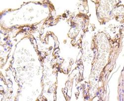
- Immunohistochemical analysis of LRP1 of paraffin-embedded Human lung tissue using a LRP1 Monoclonal antibody (Product #MA5-31959). Counter stained with hematoxylin.

- Submitted by
- Invitrogen Antibodies (provider)
- Main image
- Experimental details
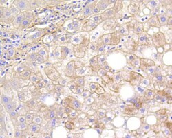
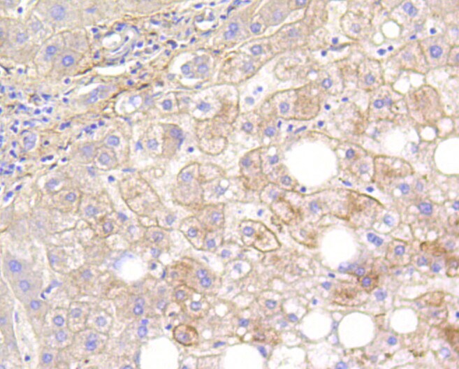
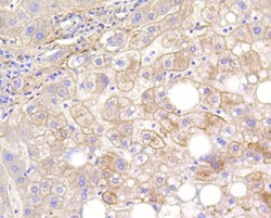
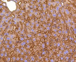
- Immunohistochemical analysis of LRP1 of paraffin-embedded Human liver tissue using a LRP1 Monoclonal antibody (Product #MA5-31959). Counter stained with hematoxylin.
- Submitted by
- Invitrogen Antibodies (provider)
- Main image
- Experimental details
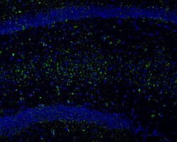
- Immunohistochemistry analysis of LRP1 in frozen mouse hippocampus. Blocked in 3% BSA for 30 minutes at room temperature, washed with PBS. Incubation was done with monoclonal LRP1 antibody (Product # MA5-31959) at a dilution of 1:100 (at 4°C), followed by Alexa Fluor 488 Goat Anti-Rabbit IgG H&L (1:200) and DAPI.

- Submitted by
- Invitrogen Antibodies (provider)
- Main image

- Experimental details
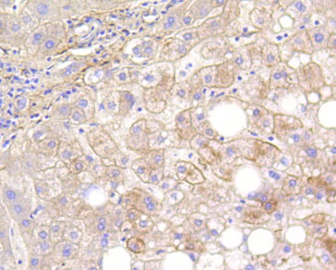
- Immunohistochemical analysis of LRP1 of paraffin-embedded Mouse liver tissue using a LRP1 Monoclonal antibody (Product #MA5-31959). Counter stained with hematoxylin.

- Submitted by
- Invitrogen Antibodies (provider)
- Main image
- Experimental details
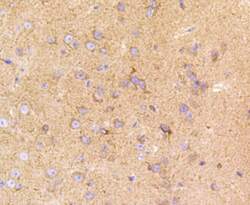
- Immunohistochemical analysis of LRP1 of paraffin-embedded Mouse brain tissue using a LRP1 Monoclonal antibody (Product #MA5-31959). Counter stained with hematoxylin.

- Submitted by
- Invitrogen Antibodies (provider)
- Main image
- Experimental details
- Immunohistochemical analysis of LRP1 of paraffin-embedded Human liver tissue using a LRP1 Monoclonal antibody (Product #MA5-31959). Counter stained with hematoxylin.
- Submitted by
- Invitrogen Antibodies (provider)
- Main image
- Experimental details
- Immunohistochemical analysis of LRP1 of paraffin-embedded Mouse liver tissue using a LRP1 Monoclonal antibody (Product #MA5-31959). Counter stained with hematoxylin.

- Submitted by
- Invitrogen Antibodies (provider)
- Main image
- Experimental details
- Immunohistochemical analysis of LRP1 of paraffin-embedded Human lung tissue using a LRP1 Monoclonal antibody (Product #MA5-31959). Counter stained with hematoxylin.

Supportive validation
- Submitted by
- Invitrogen Antibodies (provider)
- Main image

- Experimental details
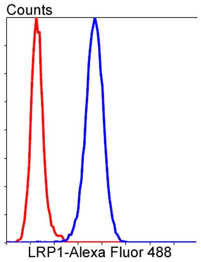
- Flow Cytometric analysis of LRP1 in Hela cells using a LRP1 Monoclonal Antibody (Product # MA5-31959) at a dilution of 1:50, as seen in blue compared with an unlabelled control (cells without incubation with primary antibody; red). Alexa Fluor 488-conjugated goat anti-rabbit IgG was used as the secondary antibody.
Supportive validation
- Submitted by
- Invitrogen Antibodies (provider)
- Main image
- Experimental details
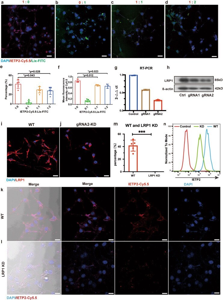
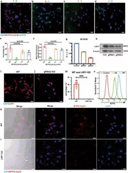
- Fig. 3 LRP1 is responsible for the endocytosis of IETP2. a - d Microscopy images showing the competing endocytosis of IETP2-Cy5.5 and Lix-FITC by HEI-OC1 cells. The endocytosis of IETP2-Cy5.5 was partially reduced by Lix-FITC. e - f Quantitative analysis of the fluorescence intensity in a - d . e Quantitative data showing the percentages of IETP2-Cy5.5-positive cells. f Quantitative data showing the mean intensity of IETP2-Cy5.5 in cells. g RT-PCR showing the knockdown (KD) efficiency of Lrp1 with two different gRNAs. h Western blot showing the expression of LRP1 after LRP1 knockdown with two different gRNAs. i , j Immunofluorescence images showing the expression of LRP1 in WT and LRP1-KD HEI-OC1 cells. k , l Microscopy images showing the endocytosis of IETP2 in WT or LRP1-KD HIE-OC1 cells. Greatly reduced endocytosis of IETP2 in LRP1-KD HEI-OC1 cells was observed. m Quantitative analysis of the images in k , l . n Flow cytometry showing the reduced endocytosis of IETP2 in LRP1-KD HEI-OC1 cells. Error bars represent the mean +- SD. N = 5 for e , f , m . N = 3 for g . * p < 0.05, *** p < 0.001. Scale bars: 20 um