Explore
 Validate
 Learn
Learn Western blot
 Immunohistochemistry
Antibody data
- Antibody Data
- Antigen structure
- References [2]
- Comments [0]
- Validations
- Immunohistochemistry [1]
- Other assay [1]
Submit
Validation data
Reference
Comment
Report error
- Product number
- PA5-28628 - Provider product page

- Provider
- Invitrogen Antibodies
- Product name
- SCRIB Polyclonal Antibody
- Antibody type
- Polyclonal
- Antigen
- Synthetic peptide
- Description
- Recommended positive controls: HepG2, NIH-3T3, JC. Predicted reactivity: Mouse (100%). Store product as a concentrated solution. Centrifuge briefly prior to opening the vial.
- Reactivity
- Human, Mouse
- Host
- Rabbit
- Isotype
- IgG
- Vial size
- 100 μL
- Concentration
- 0.13 mg/mL
- Storage
- Store at 4°C short term. For long term storage, store at -20°C, avoiding freeze/thaw cycles.
Submitted references The Dimeric Form of HPV16 E6 Is Crucial to Drive YAP/TAZ Upregulation through the Targeting of hScrib.
In-Depth Study of Transmembrane Mucins in Association with Intestinal Barrier Dysfunction During the Course of T Cell Transfer and DSS-Induced Colitis.
Messa L, Celegato M, Bertagnin C, Mercorelli B, Alvisi G, Banks L, Palù G, Loregian A
Cancers 2021 Aug 13;13(16)
Cancers 2021 Aug 13;13(16)
In-Depth Study of Transmembrane Mucins in Association with Intestinal Barrier Dysfunction During the Course of T Cell Transfer and DSS-Induced Colitis.
Breugelmans T, Van Spaendonk H, De Man JG, De Schepper HU, Jauregui-Amezaga A, Macken E, Lindén SK, Pintelon I, Timmermans JP, De Winter BY, Smet A
Journal of Crohn's & colitis 2020 Jul 30;14(7):974-994
Journal of Crohn's & colitis 2020 Jul 30;14(7):974-994
No comments: Submit comment
Supportive validation
- Submitted by
- Invitrogen Antibodies (provider)
- Main image
- Experimental details
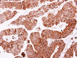
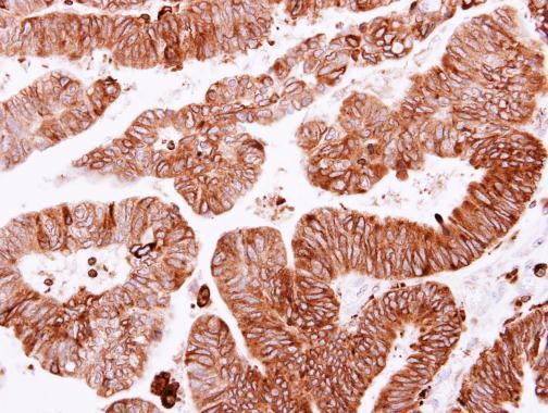
- SCRIB Polyclonal Antibody detects SCRIB protein at cytoplasm on human colon carcinoma by immunohistochemical analysis. Sample: Paraffin-embedded colon carcinoma. SCRIB Polyclonal Antibody (Product # PA5-28628) dilution: 1:250. Antigen Retrieval: EDTA based buffer, pH 8.0, 15 min.
Supportive validation
- Submitted by
- Invitrogen Antibodies (provider)
- Main image

- Experimental details
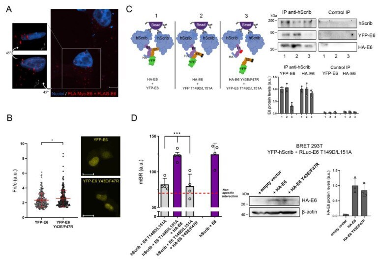
- Figure 3 The dimeric form of E6 localizes in the cytosol and takes part in the E6-hScrib complex formation. ( A ) Representative confocal image of the Myc-FLAG Proximity Ligation Assay (PLA) detecting E6 homodimerization (red fluorescence) in H1299 cells co-expressing Myc-E6 and FLAG-E6. The image represents the maximum intensity projection of a Z-stack acquired with a 2000x magnification. Scale bar: 5 mum. Left panels show the three-dimensional reconstruction of the indicated cell and both images show the same cell, with a 45 degrees rotation around the horizontal axis between the two images. Nuclei were stained with DRAQ5. Experimental controls are shown in Figure S3A,B . ( B ) Quantitative nuclear/cytoplasmic fluorescence (Fn/c) analysis of H1299 living cells overexpressing YFP-E6 or YFP-E6 Y43E/F47R. Data are presented as scatter-dot-plots of n YFP-E6 = 247 cells and n YFP-E6 Y43E/F47R = 251 cells. * p < 0.05 determined with the Mann-Whitney U test. Right panels show the representative images used for quantification. Scale bars: 20 mum. ( C ) CoIP/Western blot analysis (right) of H1299 cells overexpressing YFP-tagged and HA-tagged E6 proteins showing the binding of tagged E6 to hScrib through immunoprecipitation of endogenous hScrib. Control IP represents the extent of non-specific binding of each protein in the experiment. Left diagram provides a schematic representation of the result obtained with the experiment, with red and black circles on E6 models indicating amino