Explore
 Validate
 Learn
Learn Western blot
 Immunocytochemistry
Antibody data
- Antibody Data
- Antigen structure
- References [2]
- Comments [0]
- Validations
- Immunocytochemistry [1]
Submit
Validation data
Reference
Comment
Report error
- Product number
- HPA023557 - Provider product page

- Provider
- Atlas Antibodies
- Proper citation
- Atlas Antibodies Cat#HPA023557, RRID:AB_2671238
- Product name
- Anti-SCRIB
- Antibody type
- Polyclonal
- Description
- Polyclonal Antibody against Human SCRIB, Gene description: scribbled planar cell polarity protein, Alternative Gene Names: KIAA0147, SCRB1, Vartul, Validated applications: WB, ICC, Uniprot ID: Q14160, Storage: Store at +4°C for short term storage. Long time storage is recommended at -20°C.
- Reactivity
- Human
- Host
- Rabbit
- Conjugate
- Unconjugated
- Isotype
- IgG
- Vial size
- 100 µl
- Concentration
- 0.4 mg/ml
- Storage
- Store at +4°C for short term storage. Long time storage is recommended at -20°C.
- Handling
- The antibody solution should be gently mixed before use.
Submitted references
Polarity protein SCRIB interacts with SLC3A2 to regulate proliferation and tamoxifen resistance in ER+ breast cancer.
Mattijssen S, Kerkhofs K, Stephen J, Yang A, Han C, Tadafumi Y, Iben J, Mishra S, Sakhawala R, Ranjan A, Gowda M, Gahl W, Gu S, Malicdan M, Maraia R
2024
2024
Polarity protein SCRIB interacts with SLC3A2 to regulate proliferation and tamoxifen resistance in ER+ breast cancer.
Saito Y, Matsuda S, Ohnishi N, Endo K, Ashitani S, Ohishi M, Ueno A, Tomita M, Ueda K, Soga T, Muthuswamy SK
Communications biology 2022 May 2;5(1):403
Communications biology 2022 May 2;5(1):403
No comments: Submit comment
Supportive validation
- Submitted by
- Atlas Antibodies (provider)
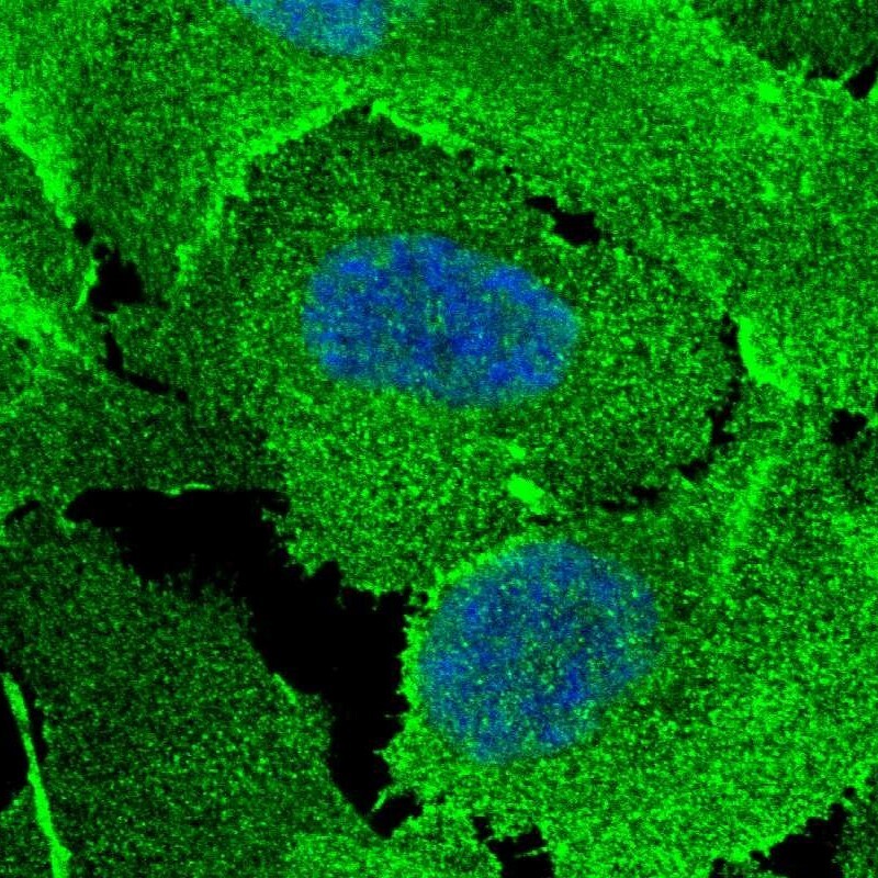
- Main image

- Experimental details
- Immunofluorescent staining of human cell line U-2 OS shows localization to nucleoplasm, plasma membrane & cell junctions.
- Sample type
- Human