Explore
 Validate
 Learn
Learn11510-100UG
antibody from Invitrogen Antibodies
Targeting: KCNQ1
JLNS1, KCNA8, KCNA9, Kv7.1, KVLQT1, LQT, LQT1
 Western blot
Antibody data
- Antibody Data
- Antigen structure
- References [0]
- Comments [0]
- Validations
- Western blot [2]
- Immunocytochemistry [1]
- Immunohistochemistry [2]
Submit
Validation data
Reference
Comment
Report error
- Product number
- 11510-100UG - Provider product page

- Provider
- Invitrogen Antibodies
- Product name
- KCNQ1 K+ Channel Monoclonal Antibody (S37A-10)
- Antibody type
- Monoclonal
- Antigen
- Other
- Reactivity
- Human, Mouse, Rat
- Host
- Mouse
- Isotype
- IgG
- Antibody clone number
- S37A-10
- Vial size
- 100 µg
- Concentration
- 1 mg/mL
- Storage
- -20° C, Avoid Freeze/Thaw Cycles
No comments: Submit comment
Supportive validation
- Submitted by
- Invitrogen Antibodies (provider)
- Main image

- Experimental details
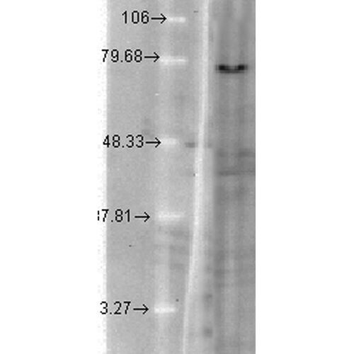
- Western Blot analysis of Hamster T-CHO cell lysate showing detection of KCNQ1 protein using Mouse Anti-KCNQ1 Monoclonal Antibody, Clone N37A/10 (11510). Load: 15 µg. Block: 1.5% BSA for 30 minutes at RT. Primary Antibody: Mouse Anti-KCNQ1 Monoclonal Antibody (11510) at 1:1000 for 2 hours at RT. Secondary Antibody: Sheep Anti-Mouse IgG: HRP for 1 hour at RT.
- Submitted by
- Invitrogen Antibodies (provider)
- Main image
- Experimental details
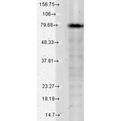
- Western Blot analysis of Human Cell lysates showing detection of KCNQ1 protein using Mouse Anti-KCNQ1 Monoclonal Antibody, Clone N37A/10 (11510). Load: 15 µg. Block: 1.5% BSA for 30 minutes at RT. Primary Antibody: Mouse Anti-KCNQ1 Monoclonal Antibody (11510) at 1:1000 for 2 hours at RT. Secondary Antibody: Sheep Anti-Mouse IgG: HRP for 1 hour at RT.

Supportive validation
- Submitted by
- Invitrogen Antibodies (provider)
- Main image
- Experimental details
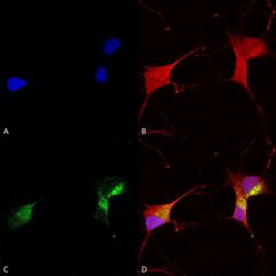
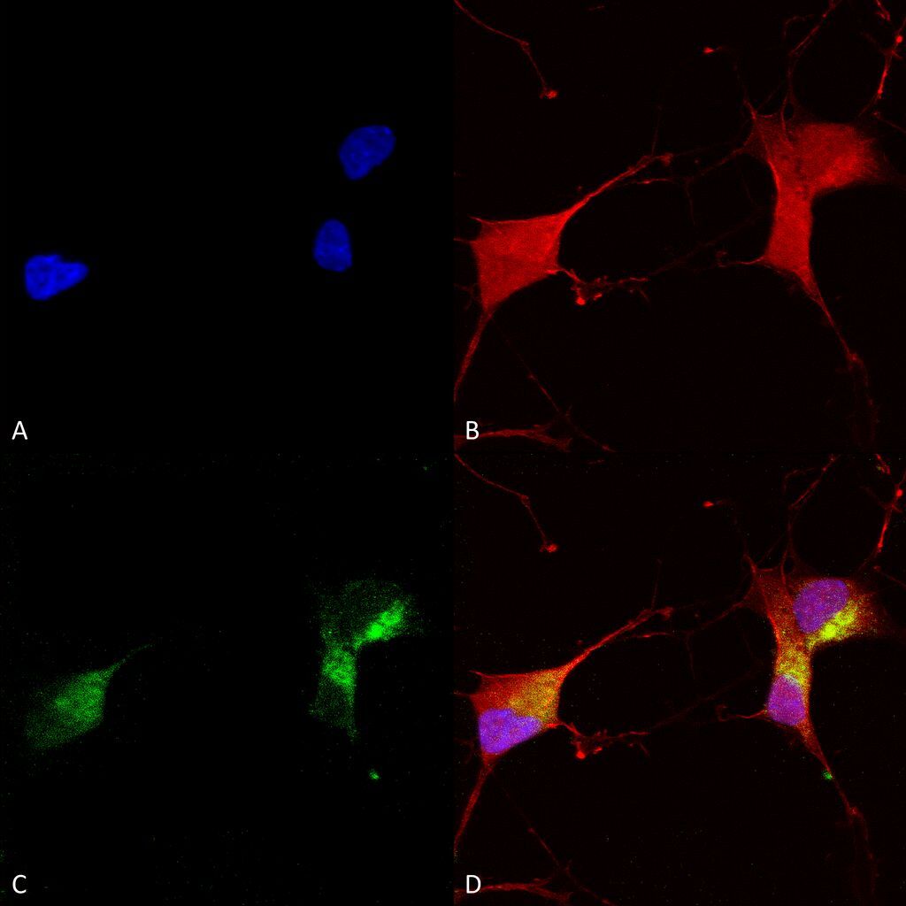
- Immunocytochemistry/Immunofluorescence analysis using Mouse Anti-KCNQ1 Monoclonal Antibody, Clone N37A/10 (11510). Tissue: Neuroblastoma cells (SH-SY5Y). Species: Human. Fixation: 4% PFA for 15 min. Primary Antibody: Mouse Anti-KCNQ1 Monoclonal Antibody (11510) at 1:100 for overnight at 4°C with slow rocking. Secondary Antibody: AlexaFluor 488 at 1:1000 for 1 hour at RT. Counterstain: Phalloidin-iFluor 647 (red) F-Actin stain; Hoechst (blue) nuclear stain at 1:800, 1.6mM for 20 min at RT. (A) Hoechst (blue) nuclear stain. (B) Phalloidin-iFluor 647 (red) F-Actin stain. (C) KCNQ1 Antibody (D) Composite.
Supportive validation
- Submitted by
- Invitrogen Antibodies (provider)
- Main image

- Experimental details
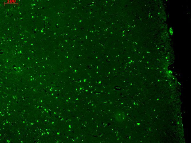
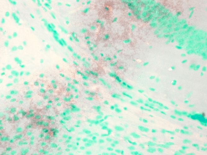
- Immunohistochemistry analysis using Mouse Anti-KCNQ1 Monoclonal Antibody, Clone N37A/10 (11510). Tissue: Brain Slice. Species: Mouse. Fixation: 10% Formalin Solution for 12-24 hours at RT. Primary Antibody: Mouse Anti-KCNQ1 Monoclonal Antibody (11510) at 1:1000 for 1 hour at RT. Secondary Antibody: HRP/DAB Detection System: Biotinylated Goat Anti-Mouse, Streptavidin Peroxidase, DAB Chromogen (brown) for 30 minutes at RT. Counterstain: Mayer Hematoxylin (purple/blue) nuclear stain at 250-500 µl for 5 minutes at RT.
- Submitted by
- Invitrogen Antibodies (provider)
- Main image
- Experimental details
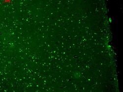
- Immunohistochemistry analysis using Mouse Anti-KCNQ1 Monoclonal Antibody, Clone N37A/10 (11510). Tissue: hippocampus. Species: Human. Fixation: Bouins Fixative and paraffin-embedded. Primary Antibody: Mouse Anti-KCNQ1 Monoclonal Antibody (11510) at 1:1000 for 1 hour at RT. Secondary Antibody: FITC Goat Anti-Mouse (green) at 1:50 for 1 hour at RT.