 Explore
 Validate
 Learn
LearnMA5-15923
antibody from Invitrogen Antibodies
Targeting: MCM2
BM28, CCNL1, cdc19, CDCL1, D3S3194, DFNA70, KIAA0030
 Western blot
Antibody data
- Antibody Data
- Antigen structure
- References [0]
- Comments [0]
- Validations
- Western blot [6]
- Immunocytochemistry [1]
- Immunohistochemistry [2]
- Flow cytometry [1]
Submit
Validation data
Reference
Comment
Report error
- Product number
- MA5-15923 - Provider product page

- Provider
- Invitrogen Antibodies
- Product name
- MCM2 Monoclonal Antibody (1E7)
- Antibody type
- Monoclonal
- Antigen
- Purifed from natural sources
- Description
- MA5-15923 targets MCM2 in FACS, IF, IHC, and WB applications and shows reactivity with Human samples.
- Antibody clone number
- 1E7
- Concentration
- Conc. Not Determined
No comments: Submit comment
Supportive validation
- Submitted by
- Invitrogen Antibodies (provider)
- Main image

- Experimental details
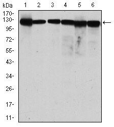
- Western blot analysis of MCM2 using MCM2 monoclonal antibody (Product # MA5-15923) in MCF-7 (1), HeLa (2), Jurkat (3), K562 (4), HEK293 (5) and HEPG2 (6) cell lysate.

- Submitted by
- Invitrogen Antibodies (provider)
- Main image

- Experimental details
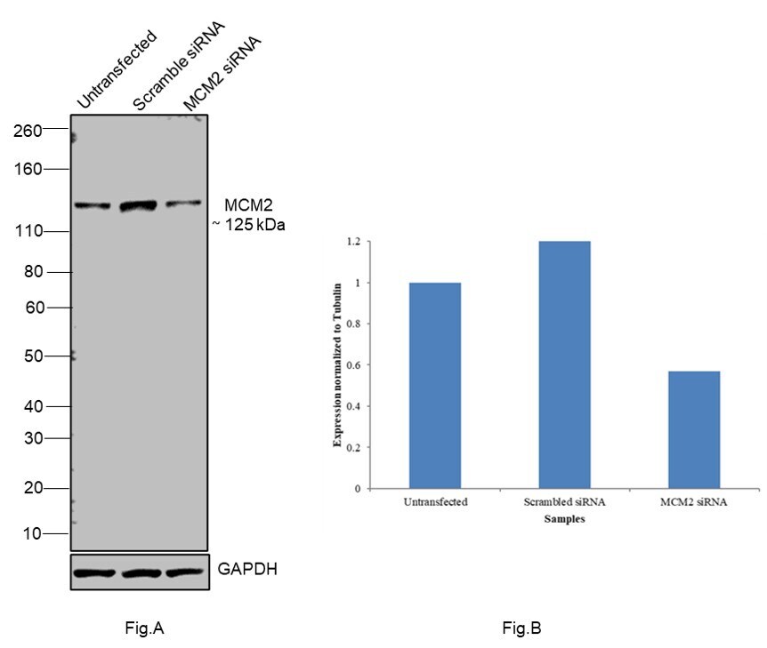
- Knockdown of MCM2 was achieved by transfecting MCF7 with MCM2 specific siRNAs (Silencer® select Product # s8586, s8587). Western blot analysis (Fig. a) was performed using Nuclear enriched extracts from the MCM2 untransfected cells (lane 1), non-targeting scrambled siRNA transfected cells (lane 2) and knockdown cells (lane 3). The blot was probed with MCM2 Monoclonal Antibody (1E7) (Product # MA5-15923, 1:1000 dilution ) and Goat anti-Mouse IgG (H+L) Superclonal™ Recombinant Secondary Antibody, HRP (Product # A28177, 1:4000 dilution). Densitometric analysis of this western blot is shown in histogram (Fig. b). Decrease in signal upon siRNA mediated knock down confirms that antibody is specific to MCM2.
- Submitted by
- Invitrogen Antibodies (provider)
- Main image

- Experimental details
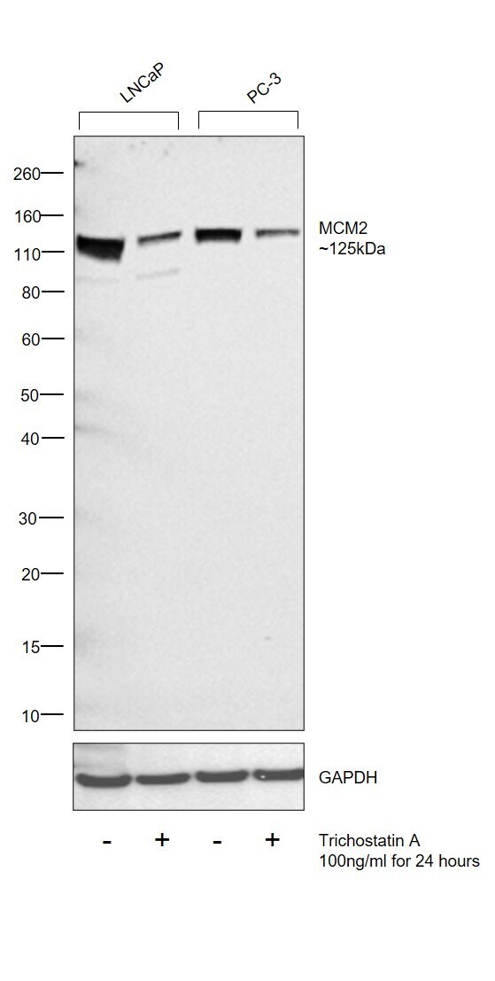
- Western blot was performed using Anti-MCM2 Monoclonal Antibody (Product # MA5-15923) and a 125 kDa band corresponding to MCM2 was observed across all cell lines. Expression of MCM2 was observed to be downregulated upon treatment of LNCaP and PC-3 cells with Trichostatin A (100 ng/mL for 24 hours) (DOI: 10.1158/0008-5472.CAN-09-4176). Modified whole cell extracts (1% SDS) (50 µg lysate) of LNCaP (Lane 1), LNCaP treated with Trichostatin A (100 ng/mL for 24 hours) (Lane 2), PC-3 (Lane 3) and PC-3 treated with Trichostatin A (100 ng/mL for 24 hours) (Lane 4) were electrophoresed using NuPAGE™ 4-12% Bis-Tris Protein Gel (Product # NP0321BOX). Resolved proteins were then transferred onto a Nitrocellulose membrane (Product # IB23001) by iBlot® 2 Dry Blotting System (Product # IB21001). The blot was probed with the primary antibody (1:2000 dilution) and detected by chemiluminescence with Goat anti-Mouse IgG (H+L), Superclonal™ Recombinant Secondary Antibody, HRP (Product # A28177, 1:4000 dilution) using the iBright FL 1000 (Product # A32752). Chemiluminescent detection was performed using Novex® ECL Reagent Kit (Product # WP20005).
- Submitted by
- Invitrogen Antibodies (provider)
- Main image

- Experimental details
- Western blot was performed using Anti-MCM2 Monoclonal Antibody (1E7)(Product # MA5-15923) and a 125kDa band corresponding to MCM2 was observed across cell lines tested. Nuclear enriched extracts (30 µg lysate) of MCF7 (Lane 1), HeLa (Lane 2), A549 (Lane 3), K-562 (Lane 4), Jurkat (Lane 5), MOLT-4 (Lane 6) were electrophoresed using NuPAGE™ 4-12% Bis-Tris Protein Gel (Product # NP0322BOX). Resolved proteins were then transferred onto a Nitrocellulose membrane (Product # IB23001) by iBlot® 2 Dry Blotting System (Product # IB21001). The blot was probed with the primary antibody (1:1000 Dilution) and detected by chemiluminescence with Goat anti-Mouse IgG (H+L) Superclonal™ Recombinant Secondary Antibody, HRP (Product # A28177,1:4000 dilution) using the iBright FL 1000 (Product # A32752). Chemiluminescent detection was performed using Novex® ECL Chemiluminescent Substrate Reagent Kit (Product # WP20005).

- Submitted by
- Invitrogen Antibodies (provider)
- Main image

- Experimental details
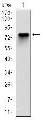
- Western blot analysis of MCM2 using a MCM2 monoclonal antibody (Product # MA5-15923) against a human MCM2 (AA: 16-232) recombinant protein.
- Submitted by
- Invitrogen Antibodies (provider)
- Main image
- Experimental details
- Western blot analysis of MCM2 using MCM2 monoclonal antibody (Product # MA5-15923) in MCF-7 (1), HeLa (2), Jurkat (3), K562 (4), HEK293 (5) and HEPG2 (6) cell lysate.

Supportive validation
- Submitted by
- Invitrogen Antibodies (provider)
- Main image

- Experimental details
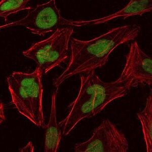
- Immunofluorescence analysis of HeLa cells using MCM2 monoclonal antibody (Product # MA5-15923) (Green). Blue: DRAQ5 fluorescent DNA dye. Red: actin filaments have been labeled with phalloidin.
Supportive validation
- Submitted by
- Invitrogen Antibodies (provider)
- Main image
- Experimental details
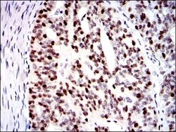
- Immunohistochemical analysis of paraffin-embedded ovarian cancer tissues using MCM2 monoclonal antibody (Product # MA5-15923) followed with DAB staining.
- Submitted by
- Invitrogen Antibodies (provider)
- Main image

- Experimental details
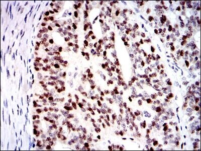
- Immunohistochemical analysis of paraffin-embedded colon cancer tissues using MCM2 monoclonal antibody (Product # MA5-15923) followed with DAB staining.
Supportive validation
- Submitted by
- Invitrogen Antibodies (provider)
- Main image

- Experimental details
- Flow cytometric analysis of Jurkat cells using MCM2 monoclonal antibody (Product # MA5-15923) (green) and negative control (red).
