Explore
 Validate
 Learn
Learn Immunohistochemistry
Antibody data
- Antibody Data
- Antigen structure
- References [1]
- Comments [0]
- Validations
- Immunohistochemistry [1]
- Other assay [1]
Submit
Validation data
Reference
Comment
Report error
- Product number
- PA5-32261 - Provider product page

- Provider
- Invitrogen Antibodies
- Product name
- Amylin Polyclonal Antibody
- Antibody type
- Polyclonal
- Antigen
- Synthetic peptide
- Description
- Heat-mediated antigen retrieval is recommended prior to staining, using a 10mM citrate buffer, pH 6.0, for 10 minutes followed by cooling at room temperature for 20 min. Following antigen retrieval, incubate samples with primary antibody for 10 min at room temperature. A suggested positive control is pancreas tissue.
- Reactivity
- Human
- Host
- Rabbit
- Isotype
- IgG
- Vial size
- 500 μL
- Storage
- -20°C, Avoid Freeze/Thaw Cycles
Submitted references Expression of the CGRP Family of Neuropeptides and their Receptors in the Trigeminal Ganglion.
Edvinsson L, Grell AS, Warfvinge K
Journal of molecular neuroscience : MN 2020 Jun;70(6):930-944
Journal of molecular neuroscience : MN 2020 Jun;70(6):930-944
No comments: Submit comment
Supportive validation
- Submitted by
- Invitrogen Antibodies (provider)
- Main image

- Experimental details
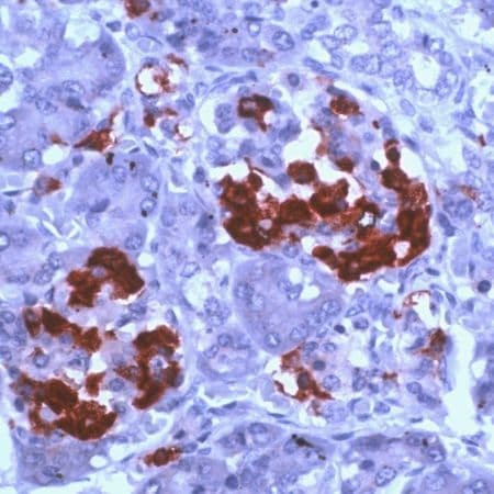
- Immunohistochemical (paraffin) analysis of Amylin Peptide using anti-Amylin Peptide Polyclonal Antibody (Product # PA5-32261) in Pancreas Cancer Tissue. The recommended dilution for this antibody in immunohistochemistry applications is 1:200.
Supportive validation
- Submitted by
- Invitrogen Antibodies (provider)
- Main image

- Experimental details
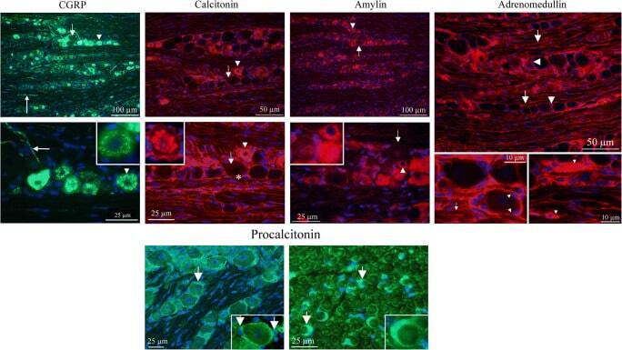
- Fig. 2 Ligand immunohistochemistry. a CGRP is expressed in a granular pattern in many neurons, mainly in small- to medium-sized neurons (arrow heads). The cellular CGRP is expressed in vesicles (insert). In addition, pearl-like CGRP immunoreactivity was detected in fibers that are of the C-type of sensory unmyelinated nerves (long arrows). The myelinated fibers do not contain CGRP. Short arrow points at a large negative neuron. CT immunoreactivity displayed a similar pattern as for CGRP; granular staining of small- to medium-sized neurons (arrow heads, insert) and pearl-like staining of fibers. Also, SGCs were CT immunoreactive (asterisks). Arrows point at a large negative neuron. AMY was exclusively expressed in the neurons, mainly small to medium sized (arrow heads). In some of the cells, the expression was granular, but in others, a general cytoplasmic immunoreactivity. Arrows point at a large negative neuron. Insert shows three different cells: one negative and two amylin immunoreactive. AM was expressed in the glial cells, both the SGCs (arrow heads) and cells enveloping the neuronal processes (arrows), probably myelinating cells. b Pro-CT was expressed exclusively in the glial cells. Panel to the left shows SGC staining (arrows) and panel to the right shows staining of the Schwann cells (arrows and insert) in a perpendicular cut of the trigeminal nerve