Explore
 Validate
 Learn
Learn Western blot
Antibody data
- Antibody Data
- Antigen structure
- References [0]
- Comments [0]
- Validations
- Western blot [3]
- Immunocytochemistry [1]
- Immunohistochemistry [2]
Submit
Validation data
Reference
Comment
Report error
- Product number
- MA5-56867 - Provider product page

- Provider
- Invitrogen Antibodies
- Product name
- RPS26 Recombinant Rabbit Monoclonal Antibody (HL3013)
- Antibody type
- Monoclonal
- Antigen
- Synthetic peptide
- Reactivity
- Human, Mouse, Rat
- Host
- Rabbit
- Isotype
- IgG
- Antibody clone number
- HL3013
- Vial size
- 100 μL
- Concentration
- 1 mg/mL
- Storage
- -20°C, Avoid Freeze/Thaw Cycles
No comments: Submit comment
Supportive validation
- Submitted by
- Invitrogen Antibodies (provider)
- Main image
- Experimental details
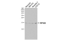
- Western Blot analysis of RPS26 using RPS26 Recombinant Monoclonal Antibody (Product # MA5-56867). Neuro2A, NIH-3T3, Raw264.7, and C2C12 whole cell extracts (30 μg) were separated by 15% SDS-PAGE, and the membrane was blotted with RPS26 antibody diluted at 1:1,000. A HRP-conjugated anti-rabbit IgG antibody was used to detect the primary antibody.
- Submitted by
- Invitrogen Antibodies (provider)
- Main image
- Experimental details
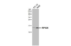
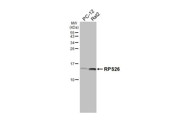
- Western Blot analysis of RPS26 using RPS26 Recombinant Monoclonal Antibody (Product # MA5-56867). PC-12 and Rat2 whole cell extracts (30 μg) were separated by 15% SDS-PAGE, and the membrane was blotted with RPS26 antibody diluted at 1:1,000. A HRP-conjugated anti-rabbit IgG antibody was used to detect the primary antibody.
- Submitted by
- Invitrogen Antibodies (provider)
- Main image
- Experimental details
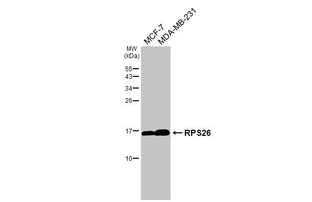
- Western Blot analysis of RPS26 using RPS26 Recombinant Monoclonal Antibody (Product # MA5-56867). MCF-7 and MDA-MB-231 whole cell extracts (30 μg) were separated by 15% SDS-PAGE, and the membrane was blotted with RPS26 antibody diluted at 1:1,000. A HRP-conjugated anti-rabbit IgG antibody was used to detect the primary antibody, and the signal was developed with Trident femto Western HRP Substrate.
Supportive validation
- Submitted by
- Invitrogen Antibodies (provider)
- Main image
- Experimental details
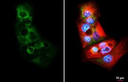
- Immunocytochemistry analysis of RPS26 using RPS26 Recombinant Monoclonal Antibody (Product # MA5-56867) in HeLa cells fixed in 4% paraformaldehyde at RT for 15 min. RPS26 antibody diluted at 1:500. Red: alpha Tubulin, a cytoskeleton marker, stained by alpha Tubulin antibody (Product # MA5-31466) Blue: Fluoroshield with DAPI.

Supportive validation
- Submitted by
- Invitrogen Antibodies (provider)
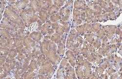
- Main image

- Experimental details
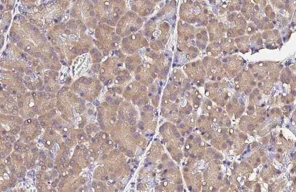
- Immunohistochemistry (Paraffin) analysis of RPS26 stained by RPS26 Recombinant Monoclonal Antibody (Product # MA5-56867). RPS26 antibody detects RPS26 protein on Paraffin-embedded rat pancreas diluted at 1:100. Antigen Retrieval: Citrate buffer, pH 6.0, 15 min.

- Submitted by
- Invitrogen Antibodies (provider)
- Main image
- Experimental details
- Immunohistochemistry (Paraffin) analysis of RPS26 stained by RPS26 Recombinant Monoclonal Antibody (Product # MA5-56867). RPS26 antibody detects RPS26 protein on Paraffin-embedded rat pancreas diluted at 1:100. Antigen Retrieval: Citrate buffer, pH 6.0, 15 min.