 Explore
 Validate
 Learn
Learn Western blot
 Immunocytochemistry
Antibody data
- Antibody Data
- Antigen structure
- References [2]
- Comments [0]
- Validations
- Western blot [3]
- Immunohistochemistry [3]
Submit
Validation data
Reference
Comment
Report error
- Product number
- NBP1-80289 - Provider product page

- Provider
- Novus Biologicals
- Proper citation
- Novus Cat#NBP1-80289, RRID:AB_11011860
- Product name
- Rabbit Polyclonal TGF-beta 1 Antibody
- Antibody type
- Polyclonal
- Description
- Immunogen affinity purified.
- Reactivity
- Human, Mouse, Porcine
- Host
- Rabbit
- Isotype
- IgG
- Vial size
- 100 ul
- Storage
- Store at -20C. Avoid freeze-thaw cycles.
Submitted references TGF-β promotes fibrosis after severe acute kidney injury by enhancing renal macrophage infiltration.
Primary proteasome inhibition results in cardiac dysfunction.
Chung S, Overstreet JM, Li Y, Wang Y, Niu A, Wang S, Fan X, Sasaki K, Jin GN, Khodo SN, Gewin L, Zhang MZ, Harris RC
JCI insight 2018 Nov 2;3(21)
JCI insight 2018 Nov 2;3(21)
Primary proteasome inhibition results in cardiac dysfunction.
Herrmann J, Wohlert C, Saguner AM, Flores A, Nesbitt LL, Chade A, Lerman LO, Lerman A
European journal of heart failure 2013 Jun;15(6):614-23
European journal of heart failure 2013 Jun;15(6):614-23
No comments: Submit comment
Supportive validation
- Submitted by
- Novus Biologicals (provider)
- Main image
- Experimental details
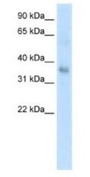
- Western Blot: TGF-beta 1 Antibody [NBP1-80289] - Antibody Titration: 0.2-1 ug/ml or 1:5000 to 1:1000 Dilution. SP2/0 cell lysate.

- Submitted by
- Novus Biologicals (provider)
- Main image
- Experimental details
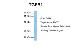
- Western Blot: TGF-beta 1 Antibody [NBP1-80289] - Sample Tissue: Human Fetal Heart. Antibody Dilution: 1.0ug/ml

- Submitted by
- Novus Biologicals (provider)
- Main image
- Experimental details
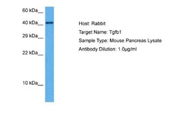
- Western Blot: TGF-beta 1 Antibody [NBP1-80289] - Sample Tissue: Mouse Pancreas. Antibody Dilution: 1ug/ml

Supportive validation
- Submitted by
- Novus Biologicals (provider)
- Main image
- Experimental details
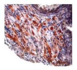
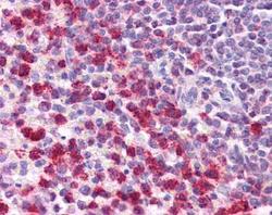
- Immunohistochemistry-Paraffin: TGF-beta 1 Antibody [NBP1-80289] - Human prostate cancer tissue was detected using RP/AEC red color stain. Working dilutions: 5-10 ug/ml.

- Submitted by
- Novus Biologicals (provider)
- Main image
- Experimental details
- Immunohistochemistry-Paraffin: TGF-beta 1 Antibody [NBP1-80289] - Human Spleen Tissue, 5 ug/ml.

- Submitted by
- Novus Biologicals (provider)
- Main image
- Experimental details
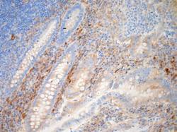
- Immunohistochemistry-Paraffin: TGF-beta 1 Antibody [NBP1-80289] - Human Appendix FFPE tissue section stained with TGF-beta 1 antibody. Image from verified customer review.
