Explore
 Validate
 Learn
Learn Western blot
 Immunocytochemistry
Antibody data
- Antibody Data
- Antigen structure
- References [0]
- Comments [0]
- Validations
- Immunocytochemistry [2]
Submit
Validation data
Reference
Comment
Report error
- Product number
- MA1-26058 - Provider product page

- Provider
- Invitrogen Antibodies
- Product name
- TGF beta-1 Monoclonal Antibody (2C5)
- Antibody type
- Monoclonal
- Antigen
- Other
- Description
- Western blotting demonstrated that this antibody reacts with the dimeric (25kDa) forms of TGF beta under both non-reducing and reducing conditions respectively.
- Reactivity
- Human
- Host
- Mouse
- Isotype
- IgG
- Antibody clone number
- 2C5
- Vial size
- 250 µg
- Concentration
- 1 mg/mL
- Storage
- Store at 4°C short term. For long term storage, store at -20°C, avoiding freeze/thaw cycles.
No comments: Submit comment
Supportive validation
- Submitted by
- Invitrogen Antibodies (provider)
- Main image

- Experimental details
- Immunofluorescence analysis of TGF beta1 (Transforming growth factor beta-1 proprotein) was performed using THP-1 and THP-1 differentiated and polarized to M2 macrophage cells. The cells were fixed with 4% paraformaldehyde for 10 minutes, permeabilized with 0.1% Triton™ X-100 for 10 minutes, and blocked with 2% BSA for 45 minutes at room temperature. The cells were labeled with TGF beta-1 Monoclonal Antibody (2C5) (Product # MA1-26058) at 1:100 in 0.1% BSA, incubated at 4 degree celsius overnight and then labeled with Donkey anti-Mouse IgG (H+L) Highly Cross-Adsorbed Secondary Antibody, Alexa Fluor Plus 488 (Product # A32766), (1:2500 dilution), for 45 minutes at room temperature (Panel a: Green). Nuclei (Panel b: Blue) were stained with ProLong™ Diamond Antifade Mountant with DAPI (Product # P36962). F-actin (Panel c: Red) was stained with Rhodamine Phalloidin (Product # R415, 1:300). Panel d represents the merged image showing increased cytoplasmic localization of TGF beta1 in M2 macrophages compared to THP-1 control cells (Panel e). Panel f represents control M2 macrophage cells with no primary antibody to assess background. The images were captured at 60X magnification.

- Submitted by
- Invitrogen Antibodies (provider)
- Main image

- Experimental details
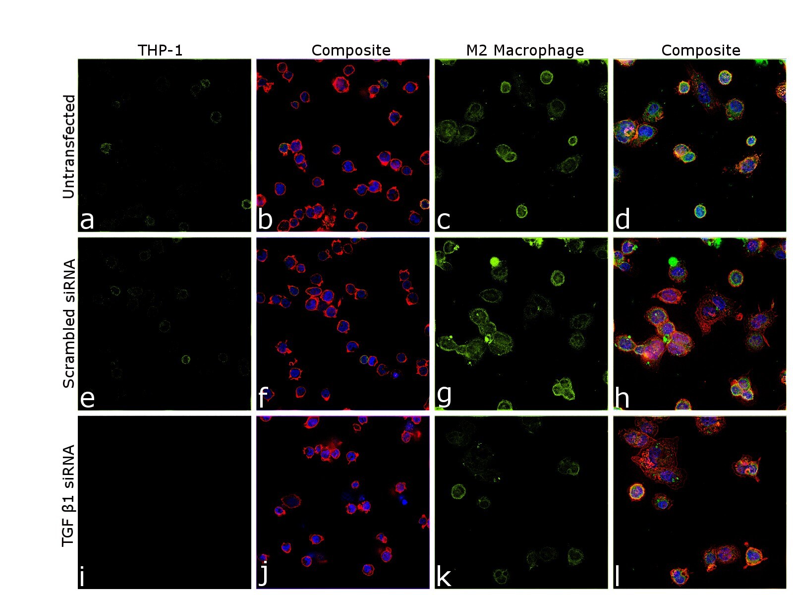
- Knockdown of TGF beta1 was achieved by transfecting THP-1 cells with TGF beta1 specific siRNA (Silencer® select Product # s14056, s14054). Immunofluorescence analysis was performed on untransfected THP1 cells (panel a,b), untransfected M2 macrophage (panel c, d), non-specific scrambled siRNA transfected THP-1 (panels e,f), non-specific scrambled siRNA transfected M2 macrophage (panels g,h), THP-1 transfected with TGF beta1 specific siRNA (panel i,j), and THP-1 transfected with TGF beta1 specific siRNA and differentiated to M2 macrophage (panels k,l) (Green). Cells were fixed, permeabilized, and labelled with TGF beta-1 Monoclonal Antibody (2C5) (Product # MA1-26058, 1:100) followed by Donkey anti-Mouse IgG (H+L) Highly Cross-Adsorbed Secondary Antibody, Alexa Fluor Plus 488 (Product # A32766), (1:2500 dilution). Nuclei (blue) were stained using ProLong™ Diamond Antifade Mountant with DAPI (Product # P36962), and Rhodamine Phalloidin (Product # R415, 1:300) was used for cytoskeletal F-actin (Blue) staining. Reduction in specific signal was observed upon siRNA mediated knockdown (panel k,l) confirming specificity of the antibody to TGF beta1. The Images were captured at 60X magnification.