 Explore
 Validate
 Learn
Learn Western blot
 ELISA
ELISAAntibody data
- Antibody Data
- Antigen structure
- References [3]
- Comments [0]
- Validations
- Western blot [2]
- Immunocytochemistry [2]
Submit
Validation data
Reference
Comment
Report error
- Product number
- MA5-18023 - Provider product page

- Provider
- Invitrogen Antibodies
- Product name
- TGF beta-1 Monoclonal Antibody (TB21)
- Antibody type
- Monoclonal
- Antigen
- Other
- Description
- This antibody reacts with the dimeric (25 kDa) and monomeric (12.5 kDa) forms of TGF-beta under both non-reducing and reducing conditions. In ELISA applications, this antibody recognizes both human platelet-derived and recombinant TGF-beta. This antibody neutralizes TGF-beta activity in vitro in an inhibition assay of CCL/64 cell growth, and neutralizes the growth promoting action of TGF-beta in the NRK-49F colony forming assay. The effect of microinjection of this antibody into one blastomere of two cell stage Xenopus embryos indicated that it was able to neutralize effectively the bioactivity of TGF-beta in vivo. In IHC applications, this antibody has been used to demonstrate TGF-beta in ovine ovarian tissue and human breast carcinoma. As a consequence of the intense staining of the erythrocytes, it is possible to locate a single cell within the ovarian stroma making it useful in locating very fine capillary networks within tissue.
- Reactivity
- Human, Mouse, Rat, Bovine, Rabbit
- Host
- Mouse
- Isotype
- IgG
- Antibody clone number
- TB21
- Vial size
- 100 µL
- Concentration
- 1 mg/mL
- Storage
- Store at 4°C short term. For long term storage, store at -20°C, avoiding freeze/thaw cycles.
Submitted references IL-10 producing CD8+ T cells in human infection with Leishmania guyanensis.
Transforming growth factor beta 1 production by CD4+ CD25+ regulatory T cells in peripheral blood mononuclear cells from healthy subjects stimulated with Leishmania guyanensis.
Tumor necrosis factor-alpha and angiostatin are mediators of endothelial cytotoxicity in bronchoalveolar lavages of patients with acute respiratory distress syndrome.
Bourreau E, Ronet C, Couppié P, Sainte-Marie D, Tacchini-Cottier F, Launois P
Microbes and infection 2007 Jul;9(8):1034-41
Microbes and infection 2007 Jul;9(8):1034-41
Transforming growth factor beta 1 production by CD4+ CD25+ regulatory T cells in peripheral blood mononuclear cells from healthy subjects stimulated with Leishmania guyanensis.
Kariminia A, Bourreau E, Pascalis H, Couppié P, Sainte-Marie D, Tacchini-Cottier F, Launois P
Infection and immunity 2005 Sep;73(9):5908-14
Infection and immunity 2005 Sep;73(9):5908-14
Tumor necrosis factor-alpha and angiostatin are mediators of endothelial cytotoxicity in bronchoalveolar lavages of patients with acute respiratory distress syndrome.
Hamacher J, Lucas R, Lijnen HR, Buschke S, Dunant Y, Wendel A, Grau GE, Suter PM, Ricou B
American journal of respiratory and critical care medicine 2002 Sep 1;166(5):651-6
American journal of respiratory and critical care medicine 2002 Sep 1;166(5):651-6
No comments: Submit comment
Supportive validation
- Submitted by
- Invitrogen Antibodies (provider)
- Main image

- Experimental details
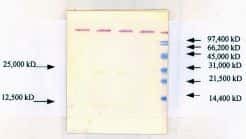
- Western blot analysis of TGF beta 1 using a TGF beta 1 monoclonal antibody (Product # MA5-18023).

- Submitted by
- Invitrogen Antibodies (provider)
- Main image

- Experimental details
- Western blot analysis of TGF beta 1 using a TGF beta 1 monoclonal antibody (Product # MA5-18023).
Supportive validation
- Submitted by
- Invitrogen Antibodies (provider)
- Main image
- Experimental details
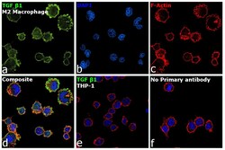
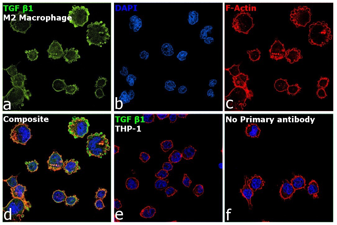
- Immunofluorescence analysis of TGF beta1 was performed using THP-1 and THP-1 differentiated and polarized to M2 macrophage cells. The cells were fixed with 4% paraformaldehyde for 10 minutes, permeabilized with 0.1% Triton™ X-100 for 10 minutes, and blocked with 2% BSA for 45 minutes at room temperature. The cells were labeled with TGF beta-1 Monoclonal Antibody (TB21) (Product # MA5-18023) at 1:100 in 0.1% BSA, incubated at 4 degree celsius overnight and then labeled with Donkey anti-Mouse IgG (H+L) Highly Cross-Adsorbed Secondary Antibody, Alexa Fluor Plus 488 (Product # A32766), (1:2500), for 45 minutes at room temperature (Panel a: Green). Nuclei (Panel b: Blue) were stained with ProLong™ Diamond Antifade Mountant with DAPI (Product # P36962). F-actin (Panel c: Red) was stained with Rhodamine Phalloidin (Product # R415, 1:300). Panel d represents the merged image showing increased expression with cytoplasmic localization of TGF beta1 in M2 macrophage compared to control THP-1 cells (panel e). Panel f represents control M2 macrophage cells with no primary antibody to assess background. The images were captured at 60X magnification.
- Submitted by
- Invitrogen Antibodies (provider)
- Main image

- Experimental details
- Knockdown of TGF beta1 was achieved by transfecting THP-1 cells with TGF beta1 specific siRNA (Silencer® select Product # s14056, s14054). Immunofluorescence analysis was performed on untransfected THP1 cells (panel a,b), untransfected M2 macrophage (panel c, d), non-specific scrambled siRNA transfected THP-1 (panels e,f), non-specific scrambled siRNA transfected M2 macrophage (panels g,h), THP-1 transfected with TGF beta1 specific siRNA (panel i,j), and THP-1 transfected with TGF beta1 specific siRNA and differentiated to M2 macrophage (panels k,l) (Green). Cells were fixed, permeabilized, and labelled with TGF beta-1 Monoclonal Antibody (TB21) (Product # MA5-18023, 1:100) followed by Donkey anti-Mouse IgG (H+L) Highly Cross-Adsorbed Secondary Antibody, Alexa Fluor Plus 488 (Product # A32766), (1:2500 dilution). Nuclei (blue) were stained using ProLong™ Diamond Antifade Mountant with DAPI (Product # P36962), and Rhodamine Phalloidin (Product # R415, 1:300) was used for cytoskeletal F-actin (Red) staining. Reduction in specific signal was observed upon siRNA mediated knockdown (panel k,l) confirming specificity of the antibody to TGF beta1. The Images were captured at 60X magnification.
