 Explore
 Validate
 Learn
Learn Western blot
 Immunocytochemistry
Antibody data
- Antibody Data
- Antigen structure
- References [0]
- Comments [0]
- Validations
- Immunocytochemistry [1]
- Immunohistochemistry [1]
- Flow cytometry [1]
- Blocking/Neutralizing [1]
Submit
Validation data
Reference
Comment
Report error
- Product number
- MAB240-100 - Provider product page

- Provider
- Novus Biologicals
- Product name
- Mouse Monoclonal TGF-beta 1 Antibody
- Antibody type
- Monoclonal
- Description
- Protein A or G purified from hybridoma culture supernatant. Detects TGF-beta 1 from human, mouse, rat, and other species in direct ELISAs and Western blots. In sandwich ELISAs, less than 2% cross-reactivity with recombinant human (rh) TGF-beta 3 and recombinant amphibian TGF-beta 5 and no cross-reactivity with recombinant porcine TGF-beta 2 or rhTGF-beta 2 is observed.
- Host
- Mouse
- Conjugate
- Unconjugated
- Isotype
- IgG
- Vial size
- 100 ug
- Storage
- Use a manual defrost freezer and avoid repeated freeze-thaw cycles. 12 months from date of receipt, -20 to -70 degreesC as supplied. 1 month, 2 to 8 degreesC under sterile conditions after reconstitution. 6 months, -20 to -70 degreesC under sterile conditions after reconstitution.
No comments: Submit comment
Supportive validation
- Submitted by
- Novus Biologicals (provider)
- Main image

- Experimental details
- TGF-beta 1 in HEK293 Human Cell Line. TGF-beta 1 was detected in immersion fixed HEK293 human embryonic kidney cell line using Mouse Anti-TGF-beta 1 Monoclonal Antibody (Catalog # MAB240) at 3 µg/mL for 3 hours at room temperature. Cells were stained using the NorthernLights™ 493-conjugated Anti-Mouse IgG Secondary Antibody (green; Catalog # NL009) and counterstained with DAPI (blue). Specific staining was localized to cytoplasm. View our protocol for Fluorescent ICC Staining of Cells on Coverslips.

Supportive validation
- Submitted by
- Novus Biologicals (provider)
- Main image
- Experimental details
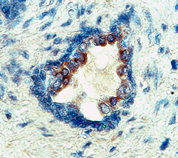
- TGF-beta 1 in Human Prostate Cancer Tissue. TGF-beta 1 was detected in immersion fixed paraffin-embedded sections of human prostate cancer tissue using Mouse Anti- TGF-beta 1 Monoclonal Antibody (Catalog # MAB240) at 25 µg/mL overnight at 4 °C. Tissue was stained using the Anti-Mouse HRP-DAB Cell & Tissue Staining Kit (brown; Catalog # CTS002) and counterstained with hematoxylin (blue). Specific labeling was localized to the cytoplasm of epithelial cells in the prostate gland. View our protocol for Chromogenic IHC Staining of Paraffin-embedded Tissue Sections.

Supportive validation
- Submitted by
- Novus Biologicals (provider)
- Main image

- Experimental details
- Detection of TGF-beta 1 in PC-3 Human Cell Line by Flow Cytometry. PC-3 human prostate cancer cell line was stained with Mouse Anti-TGF-beta 1 Monoclonal Antibody (Catalog # MAB240, filled histogram) or isotype control antibody (Catalog # MAB002, open histogram), followed by Allophycocyanin-conjugated Anti-Mouse IgG F(ab')2 Secondary Antibody (Catalog # F0101B). To facilitate intracellular staining, cells were fixed with paraformaldehyde and permeabilized with saponin.

Supportive validation
- Submitted by
- Novus Biologicals (provider)
- Main image

- Experimental details
- TGF-beta 1 Inhibition of IL-4-dependent Cell Proliferation and Neutralization by TGF-beta 1 Antibody. Recombinant Human TGF-beta 1 (Catalog # 240-B) inhibits Recombinant Mouse IL-4 (Catalog # 404-ML) induced proliferation in the HT-2 mouse T cell line in a dose-dependent manner (orange line). Inhibition of Recombinant Mouse IL-4 (7.5 ng/mL) activity elicited by Recombinant Human TGF-beta 1 (0.25 ng/mL) is neutralized (green line) by increasing concentrations of Mouse Anti-TGF-beta 1 Monoclonal Antibody (Catalog # MAB240). The ND50 is typically 0.3-1.0 µg/mL.
