Explore
 Validate
 Learn
Learn Immunocytochemistry
Antibody data
- Antibody Data
- Antigen structure
- References [5]
- Comments [0]
- Validations
- Immunocytochemistry [1]
- Other assay [9]
Submit
Validation data
Reference
Comment
Report error
- Product number
- MA5-23795 - Provider product page

- Provider
- Invitrogen Antibodies
- Product name
- TGF beta-1,2,3 Monoclonal Antibody (1D11)
- Antibody type
- Monoclonal
- Antigen
- Other
- Description
- This antibody recognizes human TGF-beta 1, TGF-beta 2, and TGF-beta 3.
- Antibody clone number
- 1D11
- Concentration
- 0.5 mg/mL
Submitted references Exposure to p40 in Early Life Prevents Intestinal Inflammation in Adulthood Through Inducing a Long-Lasting Epigenetic Imprint on TGFβ.
Dysregulation of Cytoskeleton Remodeling Drives Invasive Leading Cells Detachment.
A follicular regulatory Innate Lymphoid Cell population impairs interactions between germinal center Tfh and B cells.
Neuroprotective effects of pomegranate (Punica granatum L.) juice and seed extract in paraquat-induced mouse model of Parkinson's disease.
ΔNp63α Suppresses TGFB2 Expression and RHOA Activity to Drive Cell Proliferation in Squamous Cell Carcinomas.
Deng Y, McDonald OG, Means AL, Peek RM Jr, Washington MK, Acra SA, Polk DB, Yan F
Cellular and molecular gastroenterology and hepatology 2021;11(5):1327-1345
Cellular and molecular gastroenterology and hepatology 2021;11(5):1327-1345
Dysregulation of Cytoskeleton Remodeling Drives Invasive Leading Cells Detachment.
Peng JM, Chen WY, Cheng JH, Luo JW, Tzeng HT
Cancers 2021 Nov 11;13(22)
Cancers 2021 Nov 11;13(22)
A follicular regulatory Innate Lymphoid Cell population impairs interactions between germinal center Tfh and B cells.
O'Connor MH, Muir R, Chakhtoura M, Fang M, Moysi E, Moir S, Carey AJ, Terk A, Nichols CN, Metcalf T, Petrovas C, Cameron MJ, Tardif V, Haddad EK
Communications biology 2021 May 12;4(1):563
Communications biology 2021 May 12;4(1):563
Neuroprotective effects of pomegranate (Punica granatum L.) juice and seed extract in paraquat-induced mouse model of Parkinson's disease.
Fathy SM, El-Dash HA, Said NI
BMC complementary medicine and therapies 2021 Apr 26;21(1):130
BMC complementary medicine and therapies 2021 Apr 26;21(1):130
ΔNp63α Suppresses TGFB2 Expression and RHOA Activity to Drive Cell Proliferation in Squamous Cell Carcinomas.
Abraham CG, Ludwig MP, Andrysik Z, Pandey A, Joshi M, Galbraith MD, Sullivan KD, Espinosa JM
Cell reports 2018 Sep 18;24(12):3224-3236
Cell reports 2018 Sep 18;24(12):3224-3236
No comments: Submit comment
Supportive validation
- Submitted by
- Invitrogen Antibodies (provider)
- Main image
- Experimental details
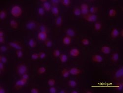
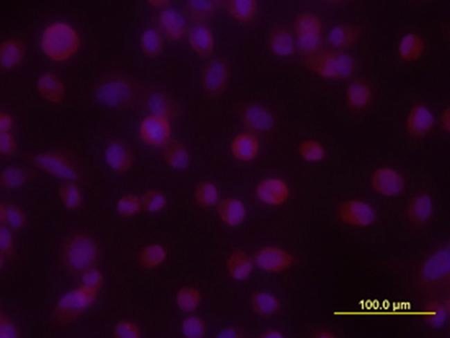
- Immunocytochemical analysis of TGF-ß1, 2, 3 was detected in immersion fixed PC-3 human prostate cancer cell line using 10 µg/mL mouse Anti-TGF-ß1, 2, 3 Monoclonal Antibody (Product # MA5-23795) for 3 hours at room temperature. Cells were stained with the 557-conjugated Anti-mouse IgG Secondary Antibody (re and counterstained with DAPI (blue).
Supportive validation
- Submitted by
- Invitrogen Antibodies (provider)
- Main image
- Experimental details
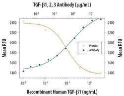
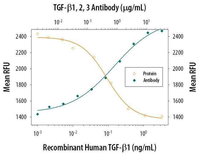
- Neutralization antibody testing demonstrates the specificity of an antibody through a correlation between antibody binding and the activity of the target. Neutralization of TGF-beta 1 is shown by increase in RFU (measure of IL-4 induced proliferation) with increasing concentrations of TGF-beta 1,2,3 monoclonal antibody (MA5-23795).
- Submitted by
- Invitrogen Antibodies (provider)
- Main image
- Experimental details
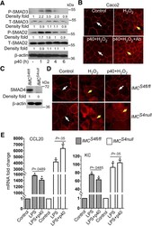
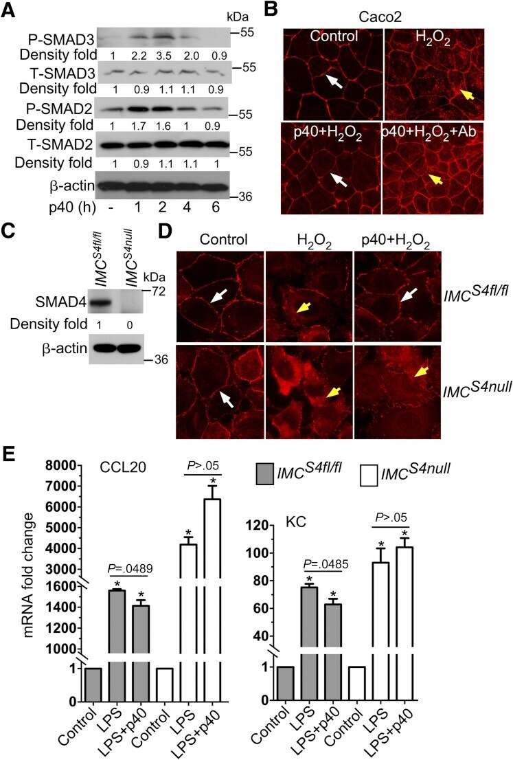
- Figure 2 p40-stimulated TGFbeta production in IECs contributes to protective epithelial cellular responses. ( A ) YAMC cells were treated with p40 (10 ng/mL) for the indicated times. Total cellular proteins were prepared for Western blot analysis. beta-actin blot was used as the protein loading control. ( B ) Caco 2 cells were treated with H 2 O 2 (20 mumol/L) for 4 hours with or without 1-hour pretreatment of p40 (10 ng/mL) and TGFbeta-neutralizing antibody (1 mug/mL). ( C ) Total cellular proteins from IMC S4fl/fl and IMC S4 cells were prepared for Western blot analysis. ( D ) IMC S4fl/fl and IMC S4 cells were treated with H 2 O 2 (20 mumol/L) for 4 hours with or without 1-hour pretreatment of p40 (10 ng/mL). The cells were fixed and immunostained to localize ZO-1 using an anti-ZO-1 antibody and a Cy3-conjugated secondary antibody (red). Nuclei were stained with 4'',6-diamidino-2-phenylindole (blue). Membrane ( white arrows ) and intracellular ( yellow arrows ) ZO-1 distribution are shown. Images were taken using a fluorescent microscope at a magnification of 40x. ( E ) Cells were treated with lipopolysaccharide (LPS) (1 mug/mL) with and without p40 (10 ng/mL) for 24 hours. RNA was isolated for RT-PCR analysis of the expression levels of indicated genes. The mRNA expression levels in the control groups were set as 1. The mRNA expression levels in treated groups were compared with the control group of the same cell line. ( A and C ) The fold changes of band density a
- Submitted by
- Invitrogen Antibodies (provider)
- Main image
- Experimental details
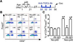
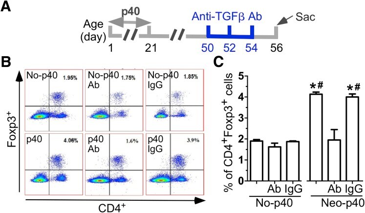
- Figure 7 Neonatal p40 supplementation-promoted Treg expansion in the lamina propria of the colon in adult mice requires sustained increase in TGFbeta production. ( A ) The treatment plan is shown. Foxp3-GFP mice were supplemented with p40 from postnatal day 2 to day 21 and received anti-TGFbeta-neutralizing antibodies or isotype-negative control antibodies (IgG) at 50 mug/d at the indicated time points. Lymphocytes were isolated from the lamina propria of the colon. CD4- and Foxp3-expressing cells were assessed using flow cytometry analysis. ( B ) Representative contour plots of Foxp3 and CD4 are shown. Numbers in quarter 2 of contour plots represent the percentages of CD4 + Foxp3 + in total lamina propria cells. ( C ) The percentages of CD4 + Foxp3 + cells in total lamina propria cells are shown. N = 3 samples. Each sample contains cells from 2 to 3 mice. * P < .05 compared with the counterpart in the no-p40 group. # P < .05 compared with the Neo-p40 group with TGFbeta antibody co-treatment. Ab, anti-TGFbeta antibody.
- Submitted by
- Invitrogen Antibodies (provider)
- Main image
- Experimental details
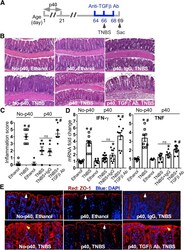
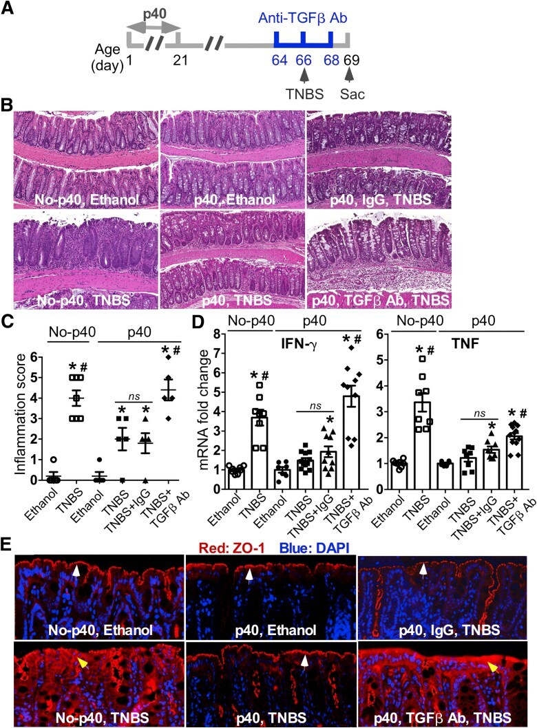
- Figure 8 Sustained TGFbeta production by neonatal p40 supplementation mediates prevention of colitis in adult mice. ( A ) The treatment plan is shown. Mice were supplemented with p40 from postnatal day 2 to day 21 and received TGFbeta-neutralizing antibodies or isotype control antibodies (IgG) at 50 mug/d, at the indicated time points. Colitis was induced by TNBS in ethanol intrarectally. Mice receiving ethanol were used as controls for TNBS treatment. Mice were killed 4 days after TNBS treatment. ( B ) Colon sections were stained with H&E for assessment of inflammation. ( C ) The inflammation scores are shown. ( D ) RNA was isolated from the colonic tissues for RT-PCR analysis of the indicated cytokine mRNA expression levels. The average cytokine mRNA expression level in the control mice of the no-p40 group was set as 1, and the mRNA expression level of each mouse was compared with this average. * P < .05 compared with the control mice in the no-p40 group. # P < .05 compared with the p40 group with TNBS or TNBS and IgG co-treatment. ( E ) Paraffin-embedded colon tissues were used to determine ZO-1 distribution by immunohistochemistry using an anti-ZO-1 antibody and a Cy3-labeled secondary antibody (red). Nuclei were stained with 4'',6-diamidino-2-phenylindole (DAPI) (blue). Slides with H&E staining and immunostaining were scanned and images were exported at 10X magnification. Membrane ( white arrowheads ) and intracellular ( yellow arrowheads ) ZO-1 distributions are shown.
- Submitted by
- Invitrogen Antibodies (provider)
- Main image
- Experimental details
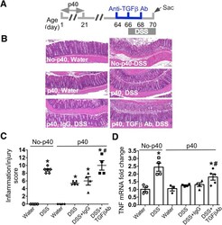
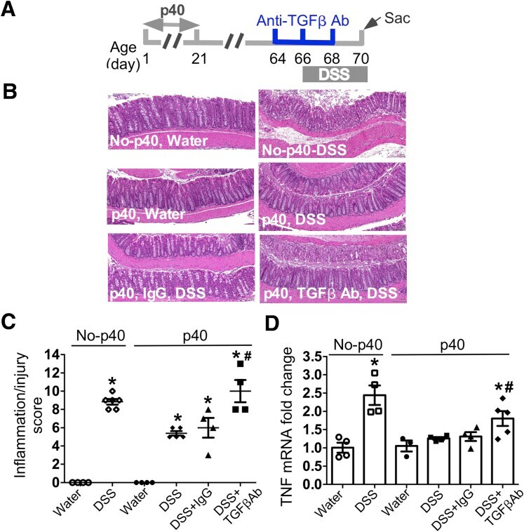
- Figure 9 Sustained TGFbeta production by neonatal p40 supplementation mediates. prevention of colitis in adult mice. ( A ) The treatment plan is shown. Mice were supplemented with p40 from postnatal day 2 to day 21 and received TGFbeta-neutralizing antibodies or isotype control antibodies (IgG) at 50 g/d, at the indicated time points. Colitis was induced by 3% DSS in drinking water for 4 days. Mice receiving water were used as controls for DSS treatment. Mice were killed at the end of DSS treatment. ( B ) Colon sections were stained with H&E for assessment of inflammation. Slides were scanned and images were exported at 10X magnification. ( C ) The inflammation/injury scores are shown. ( D ) RNA was isolated from the colonic tissues for RT-PCR analysis of the indicated cytokine mRNA expression levels. The average cytokine mRNA expression level in the control mice of the no-p40 group was set as 1, and the mRNA expression level of each mouse was compared with this average. * P < .05 compared with the control mice in the no-p40 group. # P < .05 compared with the p40 group with DSS or DSS and IgG co-treatment. Ab, antibody; Sac, sacrifice.
- Submitted by
- Invitrogen Antibodies (provider)
- Main image
- Experimental details
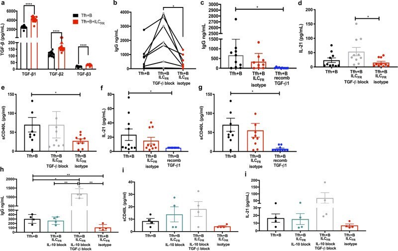
- Fig. 5 ILC FR -derived TGF-beta attenuates the interaction of GC-Tfh and GC-B cells and reverses with blockade. a Production of TGF-beta in the supernatants of 5-day co-culture: 30,000 GC-Tfh with 30,000 autologous GC-B cells (black) plus or minus addition of 1000 ILC FR (red) plus SEB measured by Luminex ( n = 12 biologically independent tonsils per group, 3 independent experiments) (Paired, two-tailed parametric t -test; **** p < 0.0001; df = 11, beta1: 95% CI: (1352 to 2048), t = 10.76, beta2: 95% CI: (32.68 to 69.68), t = 6.09, beta3: 95% CI: (9.947 to 15.43), t = 10.18; Mean +- SD). b IgG production in the supernatants of 5-day co-culture with or without blocking of TGF-beta with 1ug/mL TGF-beta neutralizing antibody (gray) or 1ug/mL mouse IgG isotype control (red) measured by ELISA ( n = 7 biologically independent tonsils per group, 3 independent experiments) (Ordinary one-way ANOVA with multiple comparisons; F = 8.380; * p = 0.0420; Mean +- SD). c IgG production in the supernatants of 5-day co-culture plus or minus 3000 pg/mL of recombinant human TGF-beta1 (blue) or 1ug/mL mouse IgG isotype control (red) measured by ELISA ( n = 10 tonsils per group, 4 independent experiments) (Ordinary one-way ANOVA with multiple comparisons; F = 3.658; * p = 0.0309; Mean +- SD). d Production of IL-21 and e sCD40L in supernatants of 5-day co-culture with or without ILC FR plus or minus TGF-beta neutralizing antibody (gray) or isotype control (red) analyzed by 28 plex Luminex ( n = 10 b
- Submitted by
- Invitrogen Antibodies (provider)
- Main image
- Experimental details
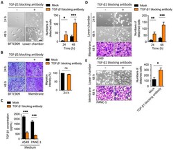
- Figure 2 Blockage of TGF-beta1 increased cell detachment after transmembrane migration. ( A ) Representative images of lower chamber and ( B ) transwell membrane demonstrated that the treatment of cancer cells with TGF-beta1-blocking antibody increased detachment in the leading cells. BFTC909 cells were seeded in the upper chamber overnight and the transwell assay was performed with or without TGF-beta1-blocking antibody in the lower chamber for 48 h. The membranes were stained by crystal violet and the cell number from membranes or the lower chamber was counted. Scale bars: 100 mum. * p < 0.05; *** p < 0.001. Data are means +- s.d. (two-way ANOVA with Tukey's multiple comparisons test and two-tailed t -test, A , B , respectively) from experiments with three replicates ( n = 3). ns, not significant. ( C ) ELISA assay showing treatment of TGF-beta1-blocking antibody reduced TGF-beta1 from the medium of the lower chamber. *** p < 0.001. Data are means +- s.d. (two-tailed t -test) from experiments with three replicates ( n = 3). ( D , E ) Representative images of lower chamber and transwell membrane demonstrated that the treatment of A549 and PANC-1 cells with TGF-beta1-blocking antibody increased detachment in the leading cells. Scale bars: 100 mum. (* p < 0.05; ** p < 0.01; *** p < 0.001). Data are means +- s.d. (two-way ANOVA with Tukey's multiple comparisons test and two-tailed t -test, D and E, respectively) from experiments with three replicates ( n = 3). Mock, mouse IgG1.

- Submitted by
- Invitrogen Antibodies (provider)
- Main image

- Experimental details
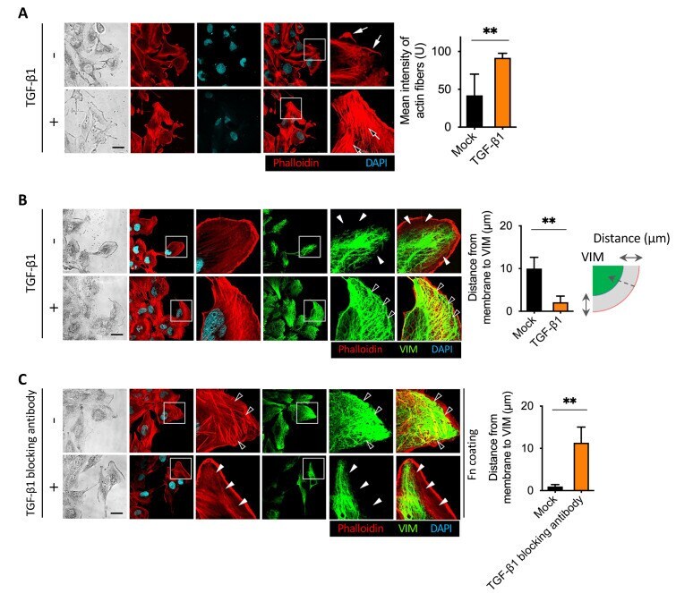
- Figure 3 TGF-beta1 induced vimentin intermediate filament networks and actin stress fibers formation. ( A ) Representative immunofluorescence assay of TGF-beta1-induced actin stress fibers formation in the leading cells. BFTC909 cells were seeded in a wound healing chamber overnight, and the migration was performed for 24 h, followed by treatment with or without TGF-beta1 ligands for another 24 h. The cells were then fixed and stained with phalloidin, and the intensity of the stress fibers was quantified and plotted. Black arrow: actin stress fibers. White arrow: lamellipodia. Scale bars: 10 mum. ** p < 0.01. Data are means +- s.d. (two-tailed t -test) from experiments with three replicates ( n = 8 fields for each experiment). ( B ) Representative images showing that TGF-beta1 promoted vimentin intermediate filament networks and actin stress fibers formation in BFTC909 cells. ** p < 0.01. Data are means +- s.d. (two-tailed t -test) from experiments with three replicates ( n = 8 fields for each experiment). ( C ) Representative images showing that TGF-beta1-blocking antibody reduced vimentin intermediate filament networks and actin stress fibers formation in BFTC909 cells. Arrowheads: the space between the intracellular membrane and vimentin. Scale bars: 10 mum. ** p < 0.01. Data are means +- s.d. (two-tailed t -test) from experiments with three replicates ( n = 8 fields for each experiment).
- Submitted by
- Invitrogen Antibodies (provider)
- Main image
- Experimental details
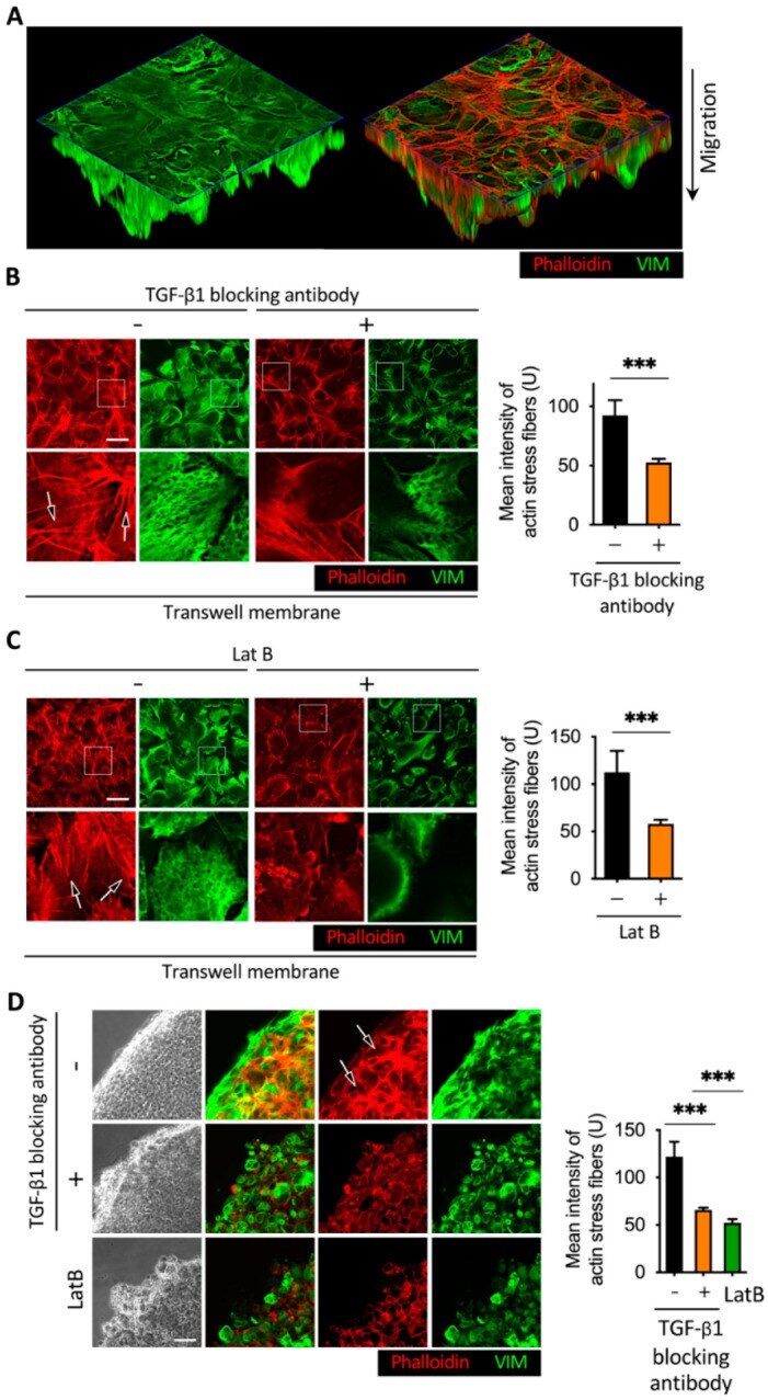
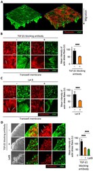
- Figure 6 Effect of TGF-beta1 neutralizing antibody and latrunculin B in the Boyden chamber and 3D sphere assay. ( A ) Immunofluorescence images of the Boyden chamber assay. ( B , C ) Images of representative immunofluorescence results revealed that the addition of TGF-beta1-blocking antibody or latrunculin B reduced actin stress fibers and vimentin intermediate filament networks. BFTC909 cells were seeded in the Boyden chamber overnight, followed by migration for 48 h. A detachment assay was performed with or without treatment of TGF-beta1 ligands or latrunculin B for another 48 h. Scale bars: 10 mum. *** p < 0.001. Data are means +- s.d. (two-tailed t test) from experiments with three replicates ( n = 3). ( D ) Treatment of TGF-beta1-blocking antibody or latrunculin B repressed actin stress fibers and vimentin intermediate filament networks in BFTC909. A three-dimensional sphere assay was performed with or without treatment of TGF-beta1-blocking antibody or latrunculin B for 48 h. Black arrow: actin stress fibers. Scale bars: 10 mum. *** p < 0.001. Data are means +- s.d. (one-way ANOVA with Dunnett's multiple comparisons test) from experiments with three replicates ( n = 3). LatB, latrunculin B.