Explore
 Validate
 Learn
Learn Western blot
Antibody data
- Antibody Data
- Antigen structure
- References [5]
- Comments [0]
- Validations
- Western blot [1]
- Immunocytochemistry [1]
- Chromatin Immunoprecipitation [1]
Submit
Validation data
Reference
Comment
Report error
- Product number
- MA5-15055 - Provider product page

- Provider
- Invitrogen Antibodies
- Product name
- c-Fos Monoclonal Antibody (T.142.5)
- Antibody type
- Monoclonal
- Antigen
- Synthetic peptide
- Description
- It is not recommended to aliquot this antibody.
- Antibody clone number
- T.142.5
- Concentration
- 108 µg/mL
Submitted references Preventive Effect of Limosilactobacillus fermentum SCHY34 on Lead Acetate-Induced Neurological Damage in SD Rats.
TIGAR deficiency enhances skeletal muscle thermogenesis by increasing neuromuscular junction cholinergic signaling.
Functional responses of the hippocampus to hyperexcitability depend on directed, neuron-specific KCNQ2 K(+) channel plasticity.
Altered chromatin landscape and enhancer engagement underlie transcriptional dysregulation in MED12 mutant uterine leiomyomas.
Environment and Behavior: Neurochemical Effects of Different Diets in the Calf Brain.
Long X, Wu H, Zhou Y, Wan Y, Kan X, Gong J, Zhao X
Frontiers in nutrition 2022;9:852012
Frontiers in nutrition 2022;9:852012
TIGAR deficiency enhances skeletal muscle thermogenesis by increasing neuromuscular junction cholinergic signaling.
Tang Y, Zong H, Kwon H, Qiu Y, Pessin JB, Wu L, Buddo KA, Boykov I, Schmidt CA, Lin CT, Neufer PD, Schwartz GJ, Kurland IJ, Pessin JE
eLife 2022 Mar 7;11
eLife 2022 Mar 7;11
Functional responses of the hippocampus to hyperexcitability depend on directed, neuron-specific KCNQ2 K(+) channel plasticity.
Carver CM, Hastings SD, Cook ME, Shapiro MS
Hippocampus 2020 May;30(5):435-455
Hippocampus 2020 May;30(5):435-455
Altered chromatin landscape and enhancer engagement underlie transcriptional dysregulation in MED12 mutant uterine leiomyomas.
Moyo MB, Parker JB, Chakravarti D
Nature communications 2020 Feb 24;11(1):1019
Nature communications 2020 Feb 24;11(1):1019
Environment and Behavior: Neurochemical Effects of Different Diets in the Calf Brain.
Peli A, Grandis A, Tassinari M, Famigli Bergamini P, Tagliavia C, Roccaro M, Bombardi C
Animals : an open access journal from MDPI 2019 Jun 14;9(6)
Animals : an open access journal from MDPI 2019 Jun 14;9(6)
No comments: Submit comment
Supportive validation
- Submitted by
- Invitrogen Antibodies (provider)
- Main image
- Experimental details
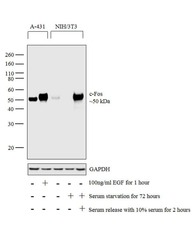
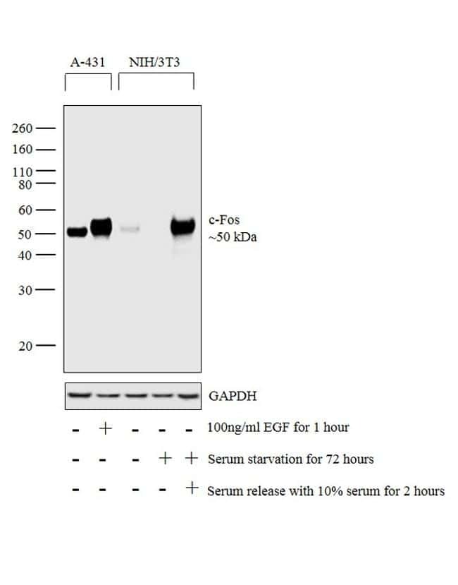
- Western blot analysis was performed on whole cell extracts (30 µg lysate) of A-431 (Lane 1), A-431 treated with EGF (100 ng/mL for 1 hour) (Lane 2), NIH/3T3 (Lane 3), NIH/3T3 Serum Starved (72 hours) (Lane 4) and NIH/3T3 Serum Starved (72 hours) followed by Serum Release (10% serum for 2 hours) (Lane 5). The blot was probed with Anti-c-Fos Monoclonal Antibody (T.142.5) (Product # MA5-15055, 1:500 dilution) and detected by chemiluminescence using Goat anti-Rabbit IgG (H+L) Superclonal™ Secondary Antibody, HRP conjugate (Product # A27036, 0.25 µg/mL, 1:4000 dilution). A 50 kDa band corresponding to c-Fos was observed across the cell lines tested and was enhanced upon treatment.
Supportive validation
- Submitted by
- Invitrogen Antibodies (provider)
- Main image

- Experimental details
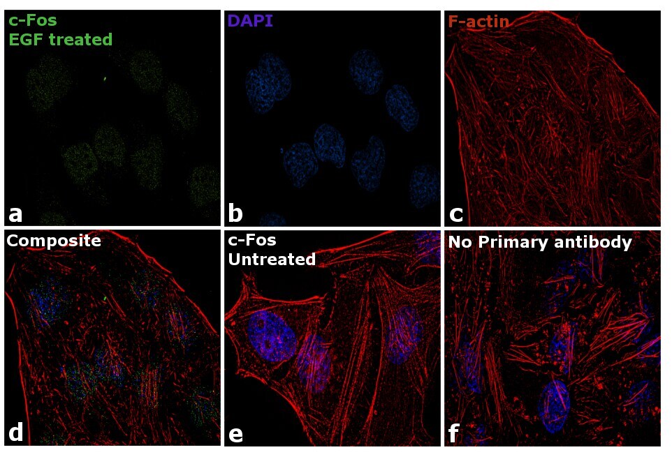
- Immunofluorescence analysis of c-Fos was performed using 70% confluent log phase HeLa cells treated with 200 ng/mL EGF for 30 min. The cells were fixed with 4% paraformaldehyde for 10 minutes, permeabilized with 0.1% Triton™ X-100 for 10 minutes, and blocked with 1% BSA for 1 hour at room temperature. The cells were labeled with c-Fos Monoclonal Antibody (T.142.5) (product # MA5-15055) at 1:250 dilution in 0.1% BSA, incubated overnight at 4 degree Celsius and then labeled with Goat anti-Rabbit IgG (H+L) Superclonal™ Secondary Antibody, Alexa Fluor® 488 conjugate (Product # A27034) at a dilution of 1:2000 for 45 minutes at room temperature (Panel a: green). Nuclei (Panel b: blue) were stained with SlowFade® Gold Antifade Mountant with DAPI (Product # S36938). F-actin (Panel c: red) was stained with Rhodamine Phalloidin (Product # R415, 1:300). Panel d represents the merged image showing nuclear localization upon EGF treatment. Panel e shows untreated cells without any staining. Panel f represents control cells with no primary antibody to assess background. The images were captured at 60X magnification.
Supportive validation
- Submitted by
- Invitrogen Antibodies (provider)
- Main image
- Experimental details
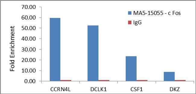
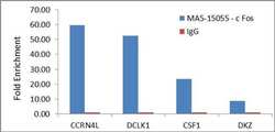
- Enrichment of endogenous c-Fos protein at specific gene loci using Anti-c-Fos Antibody: Chromatin Immunoprecipitation (ChIP) was performed using Anti-c-Fos Rabbit Monoclonal Antibody (Product # MA5-15055, 4 µg) on sheared chromatin from 2 million HeLa cells treated with EGF (200 ng/mL for 30 min) using the MAGnify ChIP system kit (Product # 49-2024). Normal Rabbit IgG was used as a negative IP control. The purified DNA was analyzed by qPCR with PCR primer pairs over CCRN4L promoter, DCLK1, CSF1 (active) and DKZ (inactive). Data is presented as fold enrichment of the antibody signal versus the negative control IgG using the comparative CT method.