Explore
 Validate
 Learn
Learn Western blot
 Immunocytochemistry
Antibody data
- Antibody Data
- Antigen structure
- References [1]
- Comments [0]
- Validations
- Western blot [2]
- Immunohistochemistry [1]
Submit
Validation data
Reference
Comment
Report error
- Product number
- NBP2-50037 - Provider product page

- Provider
- Novus Biologicals
- Proper citation
- Novus Cat#NBP2-50037, RRID:AB_2665387
- Product name
- Mouse Monoclonal c-Fos Antibody
- Antibody type
- Monoclonal
- Description
- Immunogen affinity purified.
- Reactivity
- Human, Mouse, Rat
- Host
- Mouse
- Isotype
- IgG
- Vial size
- 0.1 ml
- Concentration
- 1 mg/ml
- Storage
- Store at 4C short term. Aliquot and store at -20C long term. Avoid freeze-thaw cycles.
Submitted references Exploring mechanisms of increased cardiovascular disease risk with antipsychotic medications: Risperidone alters the cardiac proteomic signature in mice.
Beauchemin M, Geguchadze R, Guntur AR, Nevola K, Le PT, Barlow D, Rue M, Vary CPH, Lary CW, Motyl KJ, Houseknecht KL
Pharmacological research 2020 Feb;152:104589
Pharmacological research 2020 Feb;152:104589
No comments: Submit comment
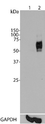
Supportive validation
- Submitted by
- Novus Biologicals (provider)
- Main image

- Experimental details
- Western Blot: c-Fos Antibody (2H2) [NBP2-50037] - Top panel: Analysis of c-Fos expression in HeLa cells using NBP2-50037. Lane 1: HeLa cells were serum-starved for 36 hours. Lane 2: Serum-starved HeLa cells were stimulated with 20% FBS (fetal bovine serum) for 2 hours. NBP2-50037 recognizes bands in the range of 50-65 kDa, which represent multiple forms of c-Fos. Serum starvation attenuates c-Fos expression, while 20% FBS strongly stimulates c-Fos expression. Bottom panel: Blot was stripped and probed with monoclonal antibody against GAPDH (NB300-221) used as loading control.
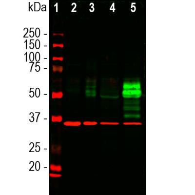
- Submitted by
- Novus Biologicals (provider)
- Main image

- Experimental details
- Western Blot: c-Fos Antibody (2H2) [NBP2-50037] - Analysis of cell lysates using mouse c-Fos mAb, dilution 1:1,000 (Green), and rabbit GAPDH pAb, dilution 1:20,000 (Red) used as a loading control. [1] protein standard (red), [2] HeLa cells in serum free media. [3] HeLa cells stimulated with 20% fetal bovine serum for 2hrs after 36hrs in serum free media. [4] rat cortical neurons. [5] rat cortical neurons treated with membrane depolarization buffer for 5hrs. Multiple bands at 50-65kDa in stimulated or treated cell lysates correspond to different forms of the c-Fos proten. The single band at 37 kDa represents GAPDH protein.
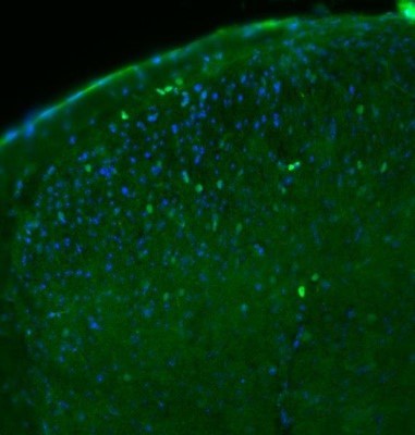
Supportive validation
- Submitted by
- Novus Biologicals (provider)
- Main image
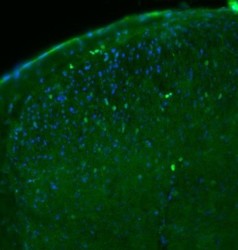
- Experimental details
- Immunohistochemistry: c-Fos Antibody (2H2) [NBP2-50037] - pAb 1:1000 (green), DAPI counterstain (blue) on 30 micron cryosection of mouse spinal cord. This image was submitted via customer Review.