Explore
 Validate
 Learn
Learn Western blot
 ELISA
ELISA Immunocytochemistry
Antibody data
- Antibody Data
- Antigen structure
- References [3]
- Comments [0]
- Validations
- Immunocytochemistry [3]
Submit
Validation data
Reference
Comment
Report error
- Product number
- MA3-218 - Provider product page

- Provider
- Invitrogen Antibodies
- Product name
- TSH Receptor Monoclonal Antibody (49)
- Antibody type
- Monoclonal
- Antigen
- Purifed from natural sources
- Description
- MA3-218 detects both glycosylated and nonglycosylated thyroid stimulating hormone receptor (TSHR) from porcine tissues as well as recombinant human TSHR. MA3-218 has been successfully used in Western blot, immunofluorescence, and ELISA procedures. By Western blot, this antibody detects a 50 kDa and a 63 kDa protein representing unglycosylated and glycosylated TSHR respectively, in transfected Sf9 cell extract. By ELISA MA3-218 has been used in TSH competitive inhibition studies. The MA3-218 antigen is purified extracellular domain of recombinant human thyrotropin receptor. This antibody recognizes an epitope between amino acid residues 277-296 of human TSHR.
- Reactivity
- Human, Porcine
- Host
- Mouse
- Isotype
- IgG
- Antibody clone number
- 49
- Vial size
- 100 μL
- Concentration
- Conc. Not Determined
- Storage
- -20°C, Avoid Freeze/Thaw Cycles
Submitted references The thyrotropin (thyroid-stimulating hormone) receptor is expressed on murine dendritic cells and on a subset of CD45RBhigh lymph node T cells: functional role for thyroid-stimulating hormone during immune activation.
Differential reactivities of recombinant glycosylated ectodomains of mouse and human thyrotropin receptors with patient autoantibodies.
Generation and characterization of monoclonal antibodies to the human thyrotropin (TSH) receptor: antibodies can bind to discrete conformational or linear epitopes and block TSH binding.
Bağriaçik EU, Klein JR
Journal of immunology (Baltimore, Md. : 1950) 2000 Jun 15;164(12):6158-65
Journal of immunology (Baltimore, Md. : 1950) 2000 Jun 15;164(12):6158-65
Differential reactivities of recombinant glycosylated ectodomains of mouse and human thyrotropin receptors with patient autoantibodies.
Patibandla SA, Seetharamaiah GS, Dallas JS, Thotakura NR, Peake RL, Prabhakar BS
Endocrinology 1997 Apr;138(4):1559-66
Endocrinology 1997 Apr;138(4):1559-66
Generation and characterization of monoclonal antibodies to the human thyrotropin (TSH) receptor: antibodies can bind to discrete conformational or linear epitopes and block TSH binding.
Seetharamaiah GS, Wagle NM, Morris JC, Prabhakar BS
Endocrinology 1995 Jul;136(7):2817-24
Endocrinology 1995 Jul;136(7):2817-24
No comments: Submit comment
Supportive validation
- Submitted by
- Invitrogen Antibodies (provider)
- Main image
- Experimental details
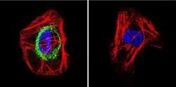
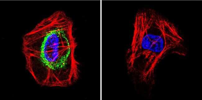
- Immunofluorescent analysis of Thyrotropin Receptor in A431 cells. Cells were grown on chamber slides and fixed with formaldehyde prior to staining. Cells were probed without (control) or with a Thyrotropin Receptor monoclonal antibody (Product # MA3-218) at a dilution of 1:200 overnight at 4 C, washed with PBS and incubated with a DyLight-488 conjugated secondary antibody (Product # 35503). Thyrotropin Receptor staining (green), F-Actin staining with Phalloidin (red) and nuclei with DAPI (blue) is shown. Images were taken at 60X magnification.
- Submitted by
- Invitrogen Antibodies (provider)
- Main image

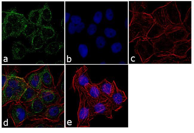
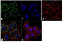
- Experimental details
- Immunofluorescence analysis of Thyrotropin Receptor was performed using 70% confluent log phase A431 cells. The cells were fixed with 4% paraformaldehyde for 10 minutes, permeabilized with 0.1% Triton™ X-100 for 10 minutes, and blocked with 1% BSA for 1 hour at room temperature. The cells were labeled with TSH Receptor (49) Mouse Monoclonal Antibody (Product # MA3-218) at 1:250 dilution in 0.1% BSA and incubated for 3 hours at room temperature and then labeled with Goat anti-Mouse IgG (H+L) Superclonal™ Secondary Antibody, Alexa Fluor® 488 conjugate (Product # A28175) at a dilution of 1:2000 for 45 minutes at room temperature (Panel a: green). Nuclei (Panel b: blue) were stained with SlowFade® Gold Antifade Mountant with DAPI (Product # S36938). F-actin (Panel c: red) was stained with Rhodamine Phalloidin (Product # R415, 1:300). Panel d represents the merged image showing membranous localization. Panel e shows the no primary antibody control. The images were captured at 60X magnification.

- Submitted by
- Invitrogen Antibodies (provider)
- Main image
- Experimental details
- Immunofluorescence analysis of Thyrotropin Receptor was performed using 70% confluent log phase A431 cells. The cells were fixed with 4% paraformaldehyde for 10 minutes, permeabilized with 0.1% Triton™ X-100 for 10 minutes, and blocked with 1% BSA for 1 hour at room temperature. The cells were labeled with TSH Receptor (49) Mouse Monoclonal Antibody (Product # MA3-218) at 1:250 dilution in 0.1% BSA and incubated for 3 hours at room temperature and then labeled with Goat anti-Mouse IgG (H+L) Superclonal™ Secondary Antibody, Alexa Fluor® 488 conjugate (Product # A28175) at a dilution of 1:2000 for 45 minutes at room temperature (Panel a: green). Nuclei (Panel b: blue) were stained with SlowFade® Gold Antifade Mountant with DAPI (Product # S36938). F-actin (Panel c: red) was stained with Rhodamine Phalloidin (Product # R415, 1:300). Panel d represents the merged image showing membranous localization. Panel e shows the no primary antibody control. The images were captured at 60X magnification.