 Explore
 Validate
 Learn
Learn Western blot
Antibody data
- Antibody Data
- Antigen structure
- References [6]
- Comments [0]
- Validations
- Western blot [1]
- Immunohistochemistry [1]
- Other assay [4]
Submit
Validation data
Reference
Comment
Report error
- Product number
- 48-3000 - Provider product page

- Provider
- Invitrogen Antibodies
- Product name
- Phospho-Connexin 43 (Ser368) Polyclonal Antibody
- Antibody type
- Polyclonal
- Antigen
- Synthetic peptide
- Description
- Aggregation: Less than 10%, as determined by HPLC.
- Reactivity
- Human, Mouse, Rat, Bovine, Canine, Chicken/Avian, Guinea Pig, Zebrafish
- Host
- Rabbit
- Isotype
- IgG
- Vial size
- 100 µL
- Storage
- Store at 4°C short term. For long term storage, store at -20°C, avoiding freeze/thaw cycles.
Submitted references Role of atrial arrhythmia and ventricular response in atrial fibrillation induced atrial remodelling.
First Responders to Hyperosmotic Stress in Murine Astrocytes: Connexin 43 Gap Junctions Are Subject to an Immediate Ultrastructural Reorganization.
Protein expression profiles in murine ventricles modeling catecholaminergic polymorphic ventricular tachycardia: effects of genotype and sex.
An N-/L-type calcium channel blocker, cilnidipine, suppresses autonomic, electrical, and structural remodelling associated with atrial fibrillation.
Subcellular reorganization and altered phosphorylation of the astrocytic gap junction protein connexin43 in human and experimental temporal lobe epilepsy.
iPS programmed without c-MYC yield proficient cardiogenesis for functional heart chimerism.
Guichard JB, Xiong F, Qi XY, L'Heureux N, Hiram R, Xiao J, Naud P, Tardif JC, Da Costa A, Nattel S
Cardiovascular research 2021 Jan 21;117(2):462-471
Cardiovascular research 2021 Jan 21;117(2):462-471
First Responders to Hyperosmotic Stress in Murine Astrocytes: Connexin 43 Gap Junctions Are Subject to an Immediate Ultrastructural Reorganization.
Beckmann A, Recktenwald J, Ferdinand A, Grißmer A, Meier C
Biology 2021 Dec 9;10(12)
Biology 2021 Dec 9;10(12)
Protein expression profiles in murine ventricles modeling catecholaminergic polymorphic ventricular tachycardia: effects of genotype and sex.
Saadeh K, Achercouk Z, Fazmin IT, Nantha Kumar N, Salvage SC, Edling CE, Huang CL, Jeevaratnam K
Annals of the New York Academy of Sciences 2020 Oct;1478(1):63-74
Annals of the New York Academy of Sciences 2020 Oct;1478(1):63-74
An N-/L-type calcium channel blocker, cilnidipine, suppresses autonomic, electrical, and structural remodelling associated with atrial fibrillation.
Tajiri K, Guichard JB, Qi X, Xiong F, Naud P, Tardif JC, Costa AD, Aonuma K, Nattel S
Cardiovascular research 2019 Dec 1;115(14):1975-1985
Cardiovascular research 2019 Dec 1;115(14):1975-1985
Subcellular reorganization and altered phosphorylation of the astrocytic gap junction protein connexin43 in human and experimental temporal lobe epilepsy.
Deshpande T, Li T, Herde MK, Becker A, Vatter H, Schwarz MK, Henneberger C, Steinhäuser C, Bedner P
Glia 2017 Nov;65(11):1809-1820
Glia 2017 Nov;65(11):1809-1820
iPS programmed without c-MYC yield proficient cardiogenesis for functional heart chimerism.
Martinez-Fernandez A, Nelson TJ, Yamada S, Reyes S, Alekseev AE, Perez-Terzic C, Ikeda Y, Terzic A
Circulation research 2009 Sep 25;105(7):648-56
Circulation research 2009 Sep 25;105(7):648-56
No comments: Submit comment
Supportive validation
- Submitted by
- Invitrogen Antibodies (provider)
- Main image

- Experimental details
- Western blot analysis of Phospho-Connexin 43 pSer368 in rat hippocampal lysate showing specific immunolabeling of the ~43 kDa Connexin 43 phosphorylated at Ser368 (Lane 1). The phosphospecificty of this labeling is shown Lane 2 (lambda-phosphatase). The blot is identical to the control except that it was incubated in lambda-phosphatase (1200 units for 30 min) before being exposed to the Phospho-Connexin 43 pSer368 polyclonal antibody (Product # 48-3000). The immunolabeling of Connexin 43 is completely eliminated by treatment with lambda-phosphatase.

Supportive validation
- Submitted by
- Invitrogen Antibodies (provider)
- Main image
- Experimental details
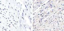
- Immunohistochemistry analysis of Phospho-Connexin 43 (pS368) showing staining in the membrane and cytoplasm of paraffin-embedded mouse heart tissue (right) compared to a negative control without primary antibody (left). To expose target proteins, antigen retrieval was performed using 10mM sodium citrate (pH 6.0), microwaved for 8-15 min. Following antigen retrieval, tissues were blocked in 3% H2O2-methanol for 15 min at room temperature, washed with ddH2O and PBS, and then probed with a Phospho-Connexin 43 (pS368) polyclonal antibody (Product # 48-3000) diluted in 3% BSA-PBS at a dilution of 1:50 overnight at 4ºC in a humidified chamber. Tissues were washed extensively in PBST and detection was performed using an HRP-conjugated secondary antibody followed by colorimetric detection using a DAB kit. Tissues were counterstained with hematoxylin and dehydrated with ethanol and xylene to prep for mounting.

Supportive validation
- Submitted by
- Invitrogen Antibodies (provider)
- Main image
- Experimental details
- NULL

- Submitted by
- Invitrogen Antibodies (provider)
- Main image

- Experimental details
- 4 Figure Expression levels obtained by densitometric analysis (upper panel) and their representative western blots and those of the housekeeping protein GAPDH, used as a loading control (lower panel) of ventricular (A) p-Cx43 and (B) TGF-beta1. Red boxes indicate female mice, and blue boxes indicate male mice. WT, wild-type; RyR2 +/+ , homozygotic RyR2 S/S .

- Submitted by
- Invitrogen Antibodies (provider)
- Main image

- Experimental details
- Figure 2 Sucrose does not affect Cx43 mRNA or protein expression. Astrocytes were treated with sucrose ( n = 3) or with PBS as control ( n = 3) for five minutes. ( A ) qPCR was performed with specific primers for Cx43 and results were normalized to 18S rRNA expression. Calculating the relative expression changes with respect to PBS-treated cells (=1) according to Pfaffl et al. revealed no changes. ( B ) SDS-PAGE of cell extracts and immunoblotting with an antibody recognizing all phosphorylation forms of Cx43 with GAPDH as a loading control were performed. Cx43 immunosignal showed unphosphorylated (P0) isoforms as well as two phosphorylated (P1/P2) isoforms. Original images are shown in Supplementary File S1 . ( C ) Quantification of signal intensities, performed on the overall immunosignal per sample, did not show any significant changes of total Cx43 protein content. Shown are means + SEM of signal intensities relative to the control. ( D ) Differential quantification of immunopositive bands P0, P1, and P2 of PBS- (black columns) and sucrose- (gray columns) treated samples. Individual comparisons of P0, P1, and P2 band intensities between groups revealed significant differences for P0 bands (** indicates p < 0.0051; uncorrected Fisher's LSD).

- Submitted by
- Invitrogen Antibodies (provider)
- Main image

- Experimental details
- Figure 4 Sucrose-treatment results in phosphorylation of Cx43 at Serine 368. Antibodies recognizing the phosphorylation of S368 were used for Cx43-immunolabeling in sucrose-treated (hyperosmolar condition) or PBS-incubated (control condition) astrocytes in immunoblots and FRIL analysis. ( A ) The immunosignal of the Cx43S368 phosphorylated isoform was enhanced in sucrose-treated astrocytes (Suc) as compared to control astrocytes (PBS). GAPDH served as a loading control. Original images are shown in Supplementary File S1 . ( B ) Quantification of n = 4 experiments confirmed a significant difference: * p < 0.05. Shown are signal intensities relative to the control (PBS), illustrated as mean + SEM. ( C ) Freeze-fractured replicas of control cells (PBS) or sucrose-stimulated cells (Sucrose) were labeled with antibodies recognizing the phosphorylation of S368. In replicas of n = 2 experiments, the number of gap junctions displaying phosphorylated Cx43S368-immunolabeling was determined. In comparison, 27 phospho (P)-Cx43S368-immunopositive gap junctions (black) and only 14 unlabeled gap junctions (gray) were found in membranes of sucrose-treated cells, whereas in membranes of control cells, only 7 gap junctions were P-Cx43S368-immunopositive (black) and 22 were found to be unlabeled (gray). ( D , E ) Examples of phospho-Cx43S368 immunolabeled gap junctions. Small and large black dots represent 6 nm and 18 nm colloidal gold, respectively, coupled to secondary antibodies. Although on
