 Explore
 Validate
 Learn
Learn Western blot
Antibody data
- Antibody Data
- Antigen structure
- References [0]
- Comments [0]
- Validations
- Western blot [4]
- Immunocytochemistry [2]
- Immunohistochemistry [1]
Submit
Validation data
Reference
Comment
Report error
- Product number
- TA328600 - Provider product page

- Provider
- OriGene
- Product name
- Rabbit Polyclonal Anti-Connexin-43
- Antibody type
- Polyclonal
- Description
- Rabbit Polyclonal Anti-Connexin-43
- Host
- Rabbit
- Conjugate
- Unconjugated
- Epitope
- GJA1
- Antibody clone number
- NULL
- Vial size
- 50 µl
- Concentration
- NULL
No comments: Submit comment
Supportive validation
- Submitted by
- OriGene (provider)
- Main image

- Experimental details
- Western blot analysis of mouse brain membranes: 1. Anti-Connexin-43 antibody(1:400). 2. Anti-Connexin-43 antibody, preincubated with the control peptide antigen.
- Validation comment
- WB

- Submitted by
- OriGene (provider)
- Main image

- Experimental details
- Western blot analysis of rat heart membranes: 1. Anti-Connexin-43 antibody(1:400). 2. Anti-Connexin-43 antibody, preincubated with the control peptide antigen.
- Validation comment
- WB

- Submitted by
- OriGene (provider)
- Main image

- Experimental details
- Western blot analysis of mouse brain membranes: 1. Anti-Connexin-43 antibody(1:400). 2. Anti-Connexin-43 antibody, preincubated with the control peptide antigen.
- Validation comment
- WB

- Submitted by
- OriGene (provider)
- Main image

- Experimental details
- Western blot analysis of rat heart membranes: 1. Anti-Connexin-43 antibody(1:400). 2. Anti-Connexin-43 antibody, preincubated with the control peptide antigen.
- Validation comment
- WB

Supportive validation
- Submitted by
- OriGene (provider)
- Main image
- Experimental details
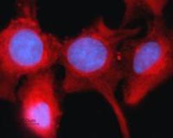
- Expression of Connexin-43 in rat intestinal epithelial IEC-6 cells . Immunocytochemical staining of fixed and permeabilized rat intestinal epithelial IEC-6 cells. Cells were stained with Anti-Connexin-43 antibody(1:200), followed by goat anti-rabbit-AlexaFluor-594 secondary antibody (red). Cell nuclei were visualized using Hoechst 33342 (blue).
- Validation comment
- IF

- Submitted by
- OriGene (provider)
- Main image

- Experimental details
- Expression of Connexin-43 in rat intestinal epithelial IEC-6 cells . Immunocytochemical staining of fixed and permeabilized rat intestinal epithelial IEC-6 cells. Cells were stained with Anti-Connexin-43 antibody(1:200), followed by goat anti-rabbit-AlexaFluor-594 secondary antibody (red). Cell nuclei were visualized using Hoechst 33342 (blue).
- Validation comment
- IF

Supportive validation
- Submitted by
- OriGene (provider)
- Main image
- Experimental details
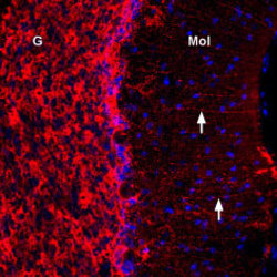
- Expression of Connexin-43 in rat cerebellum. Immunohistochemical staining of immersion-fixed, free floating rat brain frozen sections using Anti-Connexin-43 antibody(1:300). Connexin-43 staining (red) appeared in Bergmann glial fibers (arrows) in the molecular layer (MOL) and in the granule layer (G). Cell nuclei were stained with DAPI (Blue).
- Validation comment
- IHC
