Explore
 Validate
 Learn
Learn Western blot
 Immunohistochemistry
Antibody data
- Antibody Data
- Antigen structure
- References [3]
- Comments [0]
- Validations
- Immunohistochemistry [1]
- Flow cytometry [2]
- Other assay [3]
Submit
Validation data
Reference
Comment
Report error
- Product number
- 14-4759-80 - Provider product page

- Provider
- Invitrogen Antibodies
- Product name
- Connexin 43 Monoclonal Antibody (Connexin43 (1A)), eBioscience™
- Antibody type
- Monoclonal
- Antigen
- Other
- Description
- Description: The Connexin43 monoclonal antibody recognizes mouse Connexin 43. Connexins are a family of integral membrane proteins that form gap junction channels, which function to facilitate direct cell-cell communication. Gap junctions are comprised of two hemichannels, or connexons, which are made up of six connexin proteins. Over 20 connexin proteins have been identified and each combine to form either homomeric or heteromeric channels with different functional properties. Connexin 43 is expressed in many tissues, including the central nervous system, heart, and bone. Connexin 43 is the most abundant connexin found in the myocardium and is also the primary connexin expressed in astrocytes. Mutations in connexin 43 are associated with ocluodentodigital dysplasia, a rare developmental disorder characterized by craniofacial and limb abnormalities. Connexin 43 may also be a promising therapeutic target in the treatment of malignant melanomas and mammary gland tumors. The monoclonal antibody Connexin43 also recognizes human and rat connexin 43. Applications Reported: This Connexin43 antibody has been reported for use in western blotting, immunohistochemical staining of frozen tissue sections, microscopy, and immunocytochemistry. Applications Tested: This Connexin43 antibody has been tested by immunohistochemistry of frozen mouse tissue and can be used at less than or equal to 5 µg/mL. It is recommended that the antibody be carefully titrated for optimal performance in the assay of interest. Purity: Greater than 90%, as determined by SDS-PAGE. Aggregation: Less than 10%, as determined by HPLC. Filtration: 0.2 µm post-manufacturing filtered.
- Reactivity
- Human, Mouse, Rat
- Host
- Mouse
- Isotype
- IgG
- Antibody clone number
- Connexin43 (1A)
- Vial size
- 25 μg
- Concentration
- 0.5 mg/mL
- Storage
- 4°C
Submitted references A cell-penetrating peptide based on the interaction between c-Src and connexin43 reverses glioma stem cell phenotype.
Cx43 suppresses mammary tumor metastasis to the lung in a Cx43 mutant mouse model of human disease.
Oculodentodigital dysplasia connexin43 mutations result in non-functional connexin hemichannels and gap junctions in C6 glioma cells.
Gangoso E, Thirant C, Chneiweiss H, Medina JM, Tabernero A
Cell death & disease 2014 Jan 23;5(1):e1023
Cell death & disease 2014 Jan 23;5(1):e1023
Cx43 suppresses mammary tumor metastasis to the lung in a Cx43 mutant mouse model of human disease.
Plante I, Stewart MK, Barr K, Allan AL, Laird DW
Oncogene 2011 Apr 7;30(14):1681-92
Oncogene 2011 Apr 7;30(14):1681-92
Oculodentodigital dysplasia connexin43 mutations result in non-functional connexin hemichannels and gap junctions in C6 glioma cells.
Lai A, Le DN, Paznekas WA, Gifford WD, Jabs EW, Charles AC
Journal of cell science 2006 Feb 1;119(Pt 3):532-41
Journal of cell science 2006 Feb 1;119(Pt 3):532-41
No comments: Submit comment
Supportive validation
- Submitted by
- Invitrogen Antibodies (provider)
- Main image
- Experimental details
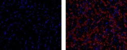
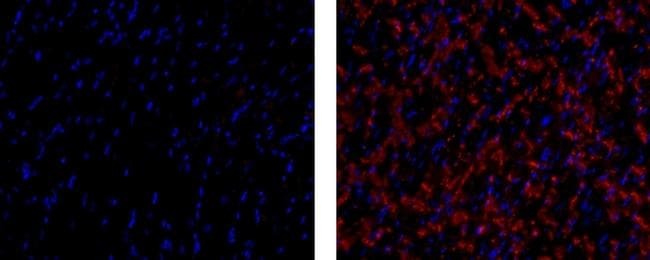
- Immunohistochemistry of frozen mouse heart using 5 µg/mL of Mouse IgG1 K Isotype Control Purified (left) or 5 µg/mL of Anti-Mouse Connexin 43 Purified (right), followed by 10 µg/mL of F (ab')2 Anti-Mouse IgG eFluor® 570. Nuclei are stained with DAPI.
Supportive validation
- Submitted by
- Invitrogen Antibodies (provider)
- Main image
- Experimental details
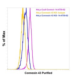
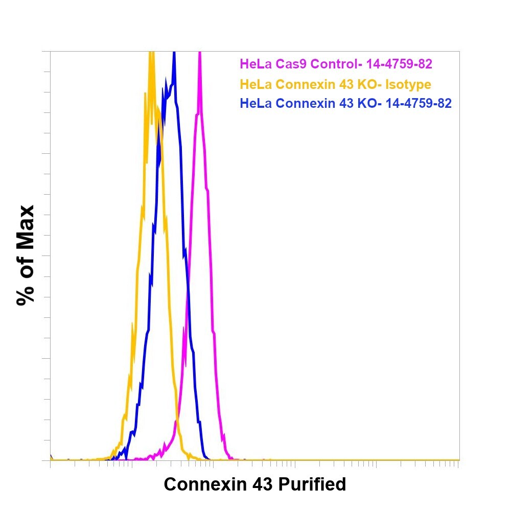
- Knockout of Connexin 43 was achieved by CRISPR-Cas9 genome editing using LentiArray™ Lentiviral sgRNA (Product # A32042, Assay ID CRISPR 888326_LV) and LentiArray Cas9 Lentivirus (Product # A32064). For Flow cytometry analysis, HeLa Connexin 43 Knock out cells were stained intracellularly using the intracellular Fixation & Permeabilization Buffer Set (Product # 88-8824-00) and protocol, with 0.5 µg Mouse IgG1 kappa Isotype Control (P3.6.2.8.1), eBioscience™ (Product # 14-4714-82, yellow histogram) or 0.5 µg Connexin 43 Monoclonal Antibody (Connexin43 (1A)), eBioscience™ (Product # 14-4759-82, blue histogram) followed by Goat anti-Mouse IgG (H+L), Superclonal™ Recombinant Secondary Antibody, Alexa Fluor™ Plus 488 (Product # A55058, 1:1000). HeLa Cas9 control cells were also stained similarly with 0.5 µg Connexin 43 Monoclonal Antibody (Connexin43 (1A)), eBioscience™ (Product # 14-4759-82, pink histogram) followed by the secondary antibody. Lossof signal was observed in the Connexin 43 KOcells stained with Connexin 43 antibody clone (Connexin43 (1A)) but not in the control Cas9cells. Viable cells were used for analysis, as determined by Fixable Viability Dye eFluor™ 780 (Product # 65-0865-18).

- Submitted by
- Invitrogen Antibodies (provider)
- Main image

- Experimental details
- Knockout of Connexin 43 was achieved by CRISPR-Cas9 genome editing using LentiArray™ Lentiviral sgRNA (Product # A32042, Assay ID CRISPR 888326_LV) and LentiArray Cas9 Lentivirus (Product # A32064). For Flow cytometry analysis, HeLa Connexin 43 Knock out cells were stained intracellularly using the intracellular Fixation & Permeabilization Buffer Set (Product # 88-8824-00) and protocol, with 0.5 µg Mouse IgG1 kappa Isotype Control (P3.6.2.8.1), eBioscience™ (Product # 14-4714-82, yellow histogram) or 0.5 µg Connexin 43 Monoclonal Antibody (Connexin43 (1A)), eBioscience™ (Product # 14-4759-82, blue histogram) followed by Goat anti-Mouse IgG (H+L), Superclonal™ Recombinant Secondary Antibody, Alexa Fluor™ Plus 488 (Product # A55058, 1:1000). HeLa Cas9 control cells were also stained similarly with 0.5 µg Connexin 43 Monoclonal Antibody (Connexin43 (1A)), eBioscience™ (Product # 14-4759-82, pink histogram) followed by the secondary antibody. Lossof signal was observed in the Connexin 43 KOcells stained with Connexin 43 antibody clone (Connexin43 (1A)) but not in the control Cas9cells. Viable cells were used for analysis, as determined by Fixable Viability Dye eFluor™ 780 (Product # 65-0865-18).
Supportive validation
- Submitted by
- Invitrogen Antibodies (provider)
- Main image
- Experimental details
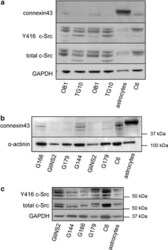
- Figure 1 Cx43 expression and c-Src activity in GSCs. ( a ) Western blot analysis of Cx43, Y416 c-Src and total c-Src in OB1 and TG10 GSCs. 58 ( b ) Western blot analysis of Cx43 expressed by G166, GliNS2, G179 and G144 GSCs characterized in Pollard et al. 32 ( c ) Western blot analysis of Y416 c-Src and total c-Src G166, GliNS2, G179 and G144 cells. Rat C6 glioma cell line and rat astrocytes from primary culture were used as controls

- Submitted by
- Invitrogen Antibodies (provider)
- Main image
- Experimental details
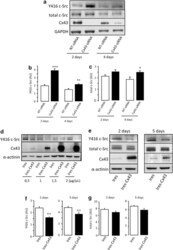
- Figure 2 Effect of Cx43 on c-Src activity. ( a - c ) G166 cells were transfected with 50 nM NT-siRNA or Cx43-siRNA. ( a ) Western blot analysis of Y416 c-Src, total c-Src, Cx43 and GAPDH 2 or 4 days after transfection. ( b ) Y416 c-Src and ( c ) total c-Src quantification. *** P

- Submitted by
- Invitrogen Antibodies (provider)
- Main image

- Experimental details
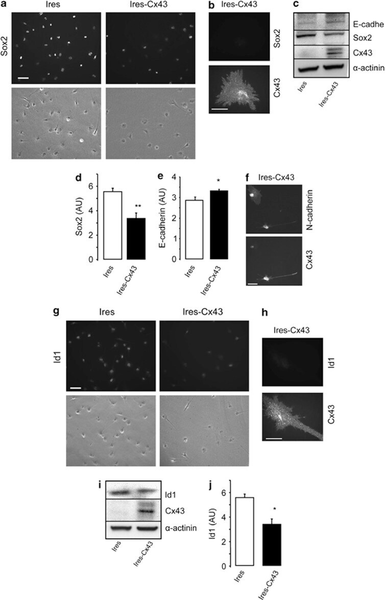
- Figure 5 Effect of restoring Cx43 on Sox2, E-cadherin, N-cadherin and Id1 expression in GSC. GliNS2 cells were transfected with the empty vector (Ires) or with the vector containing the Cx43 cDNA (Ires-Cx43). ( a ) Immunostaining and phase contrast from the same field showing the decrease in Sox2 expression in Cx43-transfected GSCs. Scale bar=20 mu m. ( b ) Double immunostaining showing the lack of Sox2 in Cx43-expressing cells. Scale bar=10 mu m. ( c ) Western blot analysis for Cx43, Sox2, E-cadherin and alpha -actinin as a loading control. ( d ) Sox2 and ( e ) E-cadherin quantification. ** P