 Explore
 Validate
 Learn
Learn Western blot
Antibody data
- Antibody Data
- Antigen structure
- References [1]
- Comments [0]
- Validations
- Western blot [1]
- Other assay [1]
Submit
Validation data
Reference
Comment
Report error
- Product number
- PA5-64640 - Provider product page

- Provider
- Invitrogen Antibodies
- Product name
- Phospho-Connexin 43 (Ser279) Polyclonal Antibody
- Antibody type
- Polyclonal
- Antigen
- Synthetic peptide
- Description
- Phospho-Connexin 43 (Ser279) Polyclonal Antibody detects endogenous levels of Connexin 43 only when phosphorylated at Ser279.
- Reactivity
- Human, Mouse, Rat
- Host
- Rabbit
- Isotype
- IgG
- Vial size
- 100 µL
- Concentration
- 1 mg/mL
- Storage
- -20°C
Submitted references Calmodulin Directly Interacts with the Cx43 Carboxyl-Terminus and Cytoplasmic Loop Containing Three ODDD-Linked Mutants (M147T, R148Q, and T154A) that Retain α-Helical Structure, but Exhibit Loss-of-Function and Cellular Trafficking Defects.
Zheng L, Chenavas S, Kieken F, Trease A, Brownell S, Anbanandam A, Sorgen PL, Spagnol G
Biomolecules 2020 Oct 17;10(10)
Biomolecules 2020 Oct 17;10(10)
No comments: Submit comment
Supportive validation
- Submitted by
- Invitrogen Antibodies (provider)
- Main image

- Experimental details
- Western blot analysis of Connexin 43 in PMA treated K562 whole cell lysates using a Phospho-Connexin 43 (Ser279) Polyclonal Antibody (Product # PA5-64640).

Supportive validation
- Submitted by
- Invitrogen Antibodies (provider)
- Main image
- Experimental details
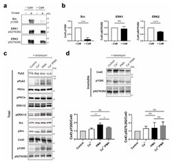
- Figure 9 Determining if the CaM interaction with Cx43CT affects MAPK and Pyk2/Src phosphorylation of the Cx43CT domain. ( a ) Src, ERK1, or ERK2 were used in an in vitro kinase assay performed with purified GST-Cx43CT 236-382 as substrate, in presence or absence of CaM. Phosphorylation was detected by Western blot using Cx43 Y265 or S279/S282 phospho-specific antibodies. ( b ) Quantification of the phosphorylation level from three independent experiments using Figures the iBright Analysis Software and analyzed in GraphPad Prism 8.0 (Student's t -test, *** p < 0.0005, **** p < 0.0001). HeLa Cx43 cells were treated with ionomycin and Ca 2+ , or PMA, or Ca 2+ /PMA for 30 min. Lysates were then subjected to Triton X-100 extraction. Total lysate ( c ) and insoluble ( d ) fractions were analyzed by Western blot. Antibodies used are labeled on the left of each panel. Quantification of the Cx43 pY265 and pS279/282 phosphorylation levels were obtained from three independent experiments using the iBright Analysis Software and analyzed in GraphPad Prism 8.0 (Ordinary one way Anova, * p < 0.05, ** p < 0.01).
