Explore
 Validate
 Learn
Learn Western blot
Antibody data
- Antibody Data
- Antigen structure
- References [0]
- Comments [0]
- Validations
- Western blot [4]
- Immunocytochemistry [2]
- Immunohistochemistry [1]
Submit
Validation data
Reference
Comment
Report error
- Product number
- ACC-201-25UL - Provider product page

- Provider
- Invitrogen Antibodies
- Product name
- Connexin-43 Polyclonal Antibody
- Antibody type
- Polyclonal
- Antigen
- Other
- Reactivity
- Human, Mouse, Rat
- Host
- Rabbit
- Isotype
- IgG
- Vial size
- 25 µL
- Concentration
- 0.8 mg/mL
- Storage
- -20° C, Avoid Freeze/Thaw Cycles
No comments: Submit comment
Supportive validation
- Submitted by
- Invitrogen Antibodies (provider)
- Main image
- Experimental details
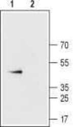
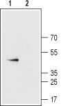
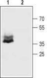
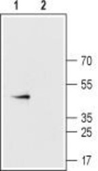
- Western blot analysis of rat heart membranes: - 1. Anti-Connexin-43 Antibody (#ACC-201), (1:400). 2. Anti-Connexin-43 Antibody , preincubated with Connexin-43 Blocking Peptide (#BLP-CC201).
- Submitted by
- Invitrogen Antibodies (provider)
- Main image
- Experimental details
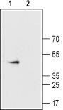
- Western blot analysis of mouse brain membranes: - 1. Anti-Connexin-43 Antibody (#ACC-201), (1:400). 2. Anti-Connexin-43 Antibody , preincubated with Connexin-43 Blocking Peptide (#BLP-CC201).

- Submitted by
- Invitrogen Antibodies (provider)
- Main image
- Experimental details
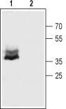
- Western blot analysis of rat heart membranes: - 1. Anti-Connexin-43 Antibody (#ACC-201), (1:400). 2. Anti-Connexin-43 Antibody , preincubated with Connexin-43 Blocking Peptide (#BLP-CC201).
- Submitted by
- Invitrogen Antibodies (provider)
- Main image

- Experimental details
- Western blot analysis of mouse brain membranes: - 1. Anti-Connexin-43 Antibody (#ACC-201), (1:400). 2. Anti-Connexin-43 Antibody , preincubated with Connexin-43 Blocking Peptide (#BLP-CC201).
Supportive validation
- Submitted by
- Invitrogen Antibodies (provider)
- Main image

- Experimental details
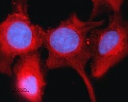
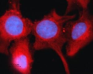
- Expression of Connexin-43 in rat intestinal epithelial IEC-6 cells - Immunocytochemical staining of fixed and permeabilized rat intestinal epithelial IEC-6 cells. Cells were stained with Anti-Connexin-43 Antibody (#ACC-201), (1:200), followed by goat Anti-rabbit-AlexaFluor-594 secondary Antibody (red). Cell nuclei were visualized using Hoechst 33342 (blue).

- Submitted by
- Invitrogen Antibodies (provider)
- Main image
- Experimental details
- Expression of Connexin-43 in rat intestinal epithelial IEC-6 cells - Immunocytochemical staining of fixed and permeabilized rat intestinal epithelial IEC-6 cells. Cells were stained with Anti-Connexin-43 Antibody (#ACC-201), (1:200), followed by goat Anti-rabbit-AlexaFluor-594 secondary Antibody (red). Cell nuclei were visualized using Hoechst 33342 (blue).
Supportive validation
- Submitted by
- Invitrogen Antibodies (provider)
- Main image
- Experimental details
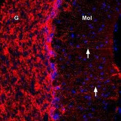
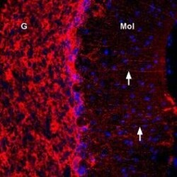
- Expression of Connexin-43 in rat cerebellum - Immunohistochemical staining of immersion-fixed, free floating rat brain frozen sections using Anti-Connexin-43 Antibody (#ACC-201), (1:300). Cx43 staining (red) appeared in Bergmann glial fibers (arrows) in the molecular layer (MOL) and in the granule layer (G). Cell nuclei were stained with DAPI (blue).