Explore
 Validate
 Learn
Learn Western blot
 Immunocytochemistry
Antibody data
- Antibody Data
- Antigen structure
- References [1]
- Comments [0]
- Validations
- Immunocytochemistry [2]
- Immunohistochemistry [1]
- Other assay [1]
Submit
Validation data
Reference
Comment
Report error
- Product number
- PA5-35054 - Provider product page

- Provider
- Invitrogen Antibodies
- Product name
- MYO6 Polyclonal Antibody
- Antibody type
- Polyclonal
- Antigen
- Synthetic peptide
- Description
- This antibody is predicted to react with mouse based on sequence homology.
- Reactivity
- Human
- Host
- Rabbit
- Isotype
- IgG
- Vial size
- 400 μL
- Concentration
- 0.5 mg/mL
- Storage
- Store at 4°C short term. For long term storage, store at -20°C, avoiding freeze/thaw cycles.
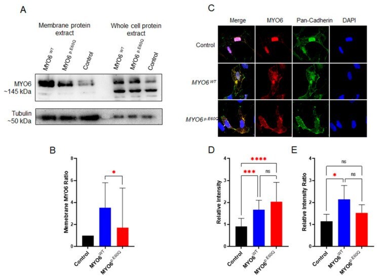
Submitted references Functional Characterization of the MYO6 Variant p.E60Q in Non-Syndromic Hearing Loss Patients.
Alkowari M, Espino-Guarch M, Daas S, Abdelrahman D, Hasan W, Krishnamoorthy N, Sathappan A, Sheehan P, Panhuys NV, The Qatar Genome Program Research Consortium, Estivill X
International journal of molecular sciences 2022 Mar 21;23(6)
International journal of molecular sciences 2022 Mar 21;23(6)
No comments: Submit comment
Supportive validation
- Submitted by
- Invitrogen Antibodies (provider)
- Main image

- Experimental details
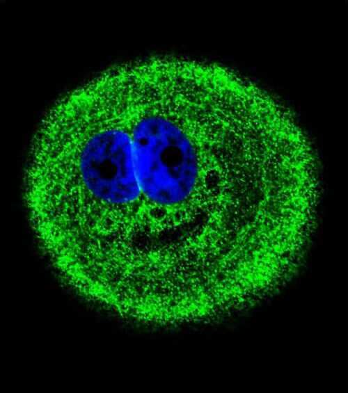
- Immunofluorescent analysis of MYO6 in MCF-7 cells using a MYO6 polyclonal antibody (Product # PA5-35054) followed by detection using a fluorescent conjugated secondary antibody (green). Nuclei were stained with Dapi (blue).
- Submitted by
- Invitrogen Antibodies (provider)
- Main image

- Experimental details
- Immunocytochemistry analysis of MYO6 in MCF-7 cells. Samples were incubated in MYO6 polyclonal antibody (Product # PA5-35054) followed by Alexa Fluor® 488-conjugated goat anti-rabbit lgG (green). DAPI was used to stain the cell nuclear (blue).

Supportive validation
- Submitted by
- Invitrogen Antibodies (provider)
- Main image
- Experimental details
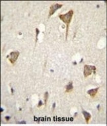
- Immunohistochemistry analysis of MYO6 in formalin fixed and paraffin embedded human brain tissue. Samples were incubated with MYO6 polyclonal antibody (Product # PA5-35054) followed by peroxidase conjugation of the secondary antibody and DAB staining. This data demonstrates the use of this antibody for immunohistochemistry. Clinical relevance has not been evaluated.

Supportive validation
- Submitted by
- Invitrogen Antibodies (provider)
- Main image
- Experimental details
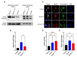
- Cellular analysis of novel MYO6 variant. ( A ) Western blot of whole-cell lysates and membrane proteins from HeL a cells transfected with plasmids carrying human MYO6 WT and MYO6 p.E60Q . ( B ) Quantification ratio of the normalized integrated density of MYO6 expression from membrane/whole proteins. ( C ) Representative immunofluorescence staining images of HeLa cells labeled by MYO6 (red), pan-cadherin (green), and DAPI (blue). ( D ) Quantification of the relative fluorescence intensity of MYO6. ( E ) Quantification of the ratio of the normalized relative fluorescence intensity of MYO6/pan-cadherin. All values are represented as the mean +- SEM from independent experiments. Statistically significant differences were assessed by ( B ) unpaired t -test, * p < 0.05 or ( D , E ) one-way ANOVA followed by Tukey's multiple comparisons, * p < 0.05; *** p = 0.0001; **** p < 0.0001.