 Explore
 Validate
 Learn
Learn Western blot
 Flow cytometry
Antibody data
- Antibody Data
- Antigen structure
- References [0]
- Comments [0]
- Validations
- Western blot [5]
- Immunocytochemistry [1]
- Immunohistochemistry [5]
Submit
Validation data
Reference
Comment
Report error
- Product number
- MA5-25561 - Provider product page

- Provider
- Invitrogen Antibodies
- Product name
- beta-4 Tubulin Monoclonal Antibody (OTI3F1)
- Antibody type
- Monoclonal
- Antigen
- Recombinant full-length protein
- Reactivity
- Human, Mouse, Rat, Canine
- Host
- Mouse
- Isotype
- IgG
- Antibody clone number
- OTI3F1
- Vial size
- 100 µL
- Concentration
- 0.57 mg/mL
- Storage
- -20° C, Avoid Freeze/Thaw Cycles
No comments: Submit comment
Supportive validation
- Submitted by
- Invitrogen Antibodies (provider)
- Main image
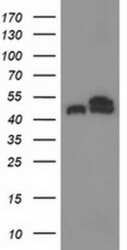
- Experimental details
- Western blot analysis of TUBB4 in HEK293T cells in untransfected (Left lane) and transfected (Right lane) samples using 5 µg per lane. The samples were separated by SDS-PAGE and probed with TUBB4 (Product # MA5-25561) monoclonal antibody.

- Submitted by
- Invitrogen Antibodies (provider)
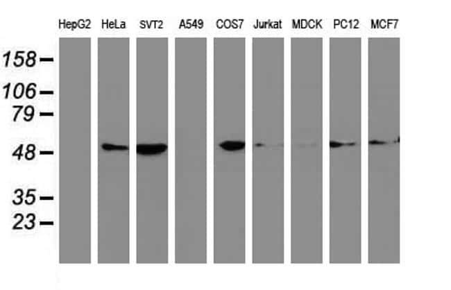
- Main image

- Experimental details
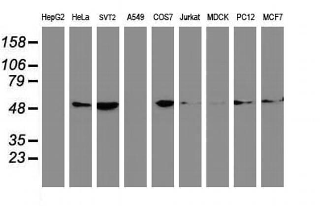
- Western blot analysis of TUBB4 in HepG2, HeLa, SVT2, A549, COS7, Jurkat, MDCK, PC12, MCF7 cells using 35 µg per lane. Samples were probed with TUBB4 (Product # MA5-25561) monoclonal antibody.
- Submitted by
- Invitrogen Antibodies (provider)
- Main image
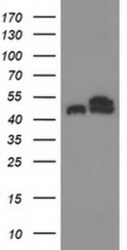
- Experimental details
- Western blot analysis of TUBB4 in HEK293T cells in untransfected (Left lane) and transfected (Right lane) samples using 5 µg per lane. The samples were separated by SDS-PAGE and probed with TUBB4 (Product # MA5-25561) monoclonal antibody.

- Submitted by
- Invitrogen Antibodies (provider)
- Main image
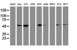
- Experimental details
- Western blot analysis of TUBB4 in HepG2, HeLa, SVT2, A549, COS7, Jurkat, MDCK, PC12, MCF7 cells using 35 µg per lane. Samples were probed with TUBB4 (Product # MA5-25561) monoclonal antibody.
- Submitted by
- Invitrogen Antibodies (provider)
- Main image
- Experimental details
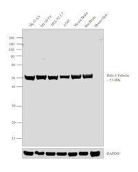
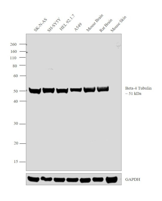
- Western blot analysis was performed on whole cell extracts (30 µg lysate) of SK-N-AS (Lane 1), SH-SY5Y (Lane 2), HEL 92.1.7 (Lane 3), A549 (Lane 4), tissue extracts of Mouse Brain (Lane 5), Rat Brain (Lane 6) and Mouse Skin (Lane 7). The blot was probed with Anti- beta-4 Tubulin Monoclonal Antibody (OTI3F1) (Product # MA5-25561, 1:500 dilution) and detected by chemiluminescence using Goat anti Mouse IgG (H+L) Superclonal™ Secondary Antibody, HRP conjugate (Product # A28177, 0.25 µg/mL, 1:4000 dilution). A 51 kDa band corresponding to beta-4 Tubulin was detected across cell lines and tissues tested except Mouse Skin which is reported to be negative for beta-4 Tubulin expression.
Supportive validation
- Submitted by
- Invitrogen Antibodies (provider)
- Main image
- Experimental details
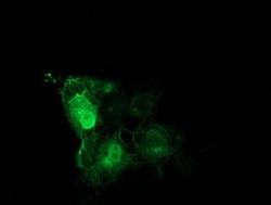
- Immunofluorescent analysis of TUBB4 in COS7 cells. Cells were transfected with a plasmid overexpressing TUBB4 and probed with a TUBB4 monoclonal antibody (Product # MA5-25561).

Supportive validation
- Submitted by
- Invitrogen Antibodies (provider)
- Main image

- Experimental details
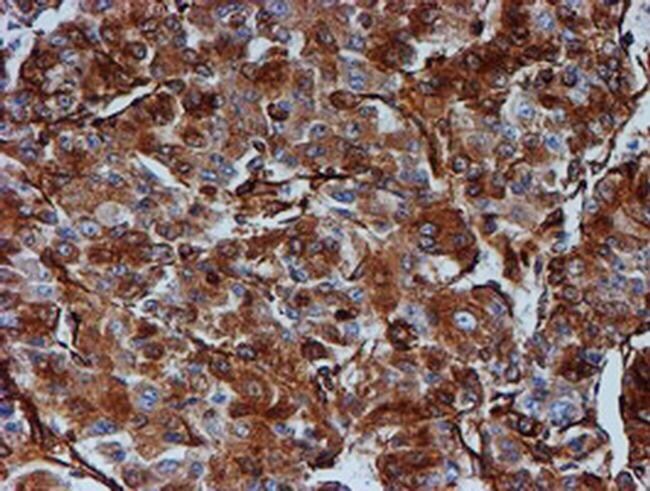
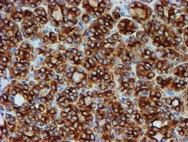
- Immunohistochemistry was performed on paraffin-embedded carcinoma of human liver tissue. To expose target proteins, 10mM citric buffer, pH6.0, 100°C for 10min was used. Following antigen retrieval, tissues were probed with a TUBB4 monoclonal antibody (Product # MA5-25561).
- Submitted by
- Invitrogen Antibodies (provider)
- Main image

- Experimental details
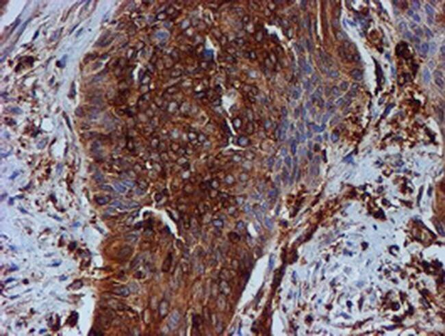
- Immunohistochemistry was performed on paraffin-embedded carcinoma of human bladder tissue. To expose target proteins, 10mM citric buffer, pH6.0, 100°C for 10min was used. Following antigen retrieval, tissues were probed with a TUBB4 monoclonal antibody (Product # MA5-25561).
- Submitted by
- Invitrogen Antibodies (provider)
- Main image
- Experimental details
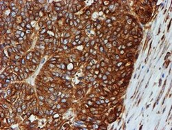
- Immunohistochemistry was performed on paraffin-embedded adenocarcinoma of human ovary tissue. To expose target proteins, 10mM citric buffer, pH6.0, 100°C for 10min was used. Following antigen retrieval, tissues were probed with a TUBB4 monoclonal antibody (Product # MA5-25561).

- Submitted by
- Invitrogen Antibodies (provider)
- Main image
- Experimental details
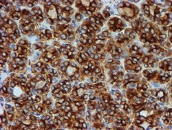
- Immunohistochemistry was performed on paraffin-embedded carcinoma of human thyroid tissue. To expose target proteins, 10mM citric buffer, pH6.0, 100°C for 10min was used. Following antigen retrieval, tissues were probed with a TUBB4 monoclonal antibody (Product # MA5-25561).
- Submitted by
- Invitrogen Antibodies (provider)
- Main image

- Experimental details
- Immunohistochemistry was performed on paraffin-embedded human tonsil tissue. To expose target proteins, 10mM citric buffer, pH6.0, 100°C for 10min was used. Following antigen retrieval, tissues were probed with a TUBB4 monoclonal antibody (Product # MA5-25561).
