Explore
 Validate
 Learn
Learn Western blot
Antibody data
- Antibody Data
- Antigen structure
- References [0]
- Comments [0]
- Validations
- Western blot [3]
- Immunocytochemistry [4]
Submit
Validation data
Reference
Comment
Report error
- Product number
- PA5-21416 - Provider product page

- Provider
- Invitrogen Antibodies
- Product name
- beta-4 Tubulin Polyclonal Antibody
- Antibody type
- Polyclonal
- Antigen
- Recombinant protein fragment
- Description
- Recommended positive controls: 293T, NIH-3T3.
- Concentration
- 0.41 mg/mL
No comments: Submit comment
Supportive validation
- Submitted by
- Invitrogen Antibodies (provider)
- Main image
- Experimental details
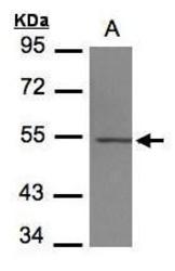
- Western Blot using beta-4 Tubulin Polyclonal Antibody (Product # PA5-21416). Sample (30 µg of whole cell lysate). Lane A: NIH-3T3. 10% SDS PAGE. beta-4 Tubulin Polyclonal Antibody (Product # PA5-21416) diluted at 1:1,000. The HRP-conjugated anti-rabbit IgG antibody was used to detect the primary antibody.

- Submitted by
- Invitrogen Antibodies (provider)
- Main image

- Experimental details
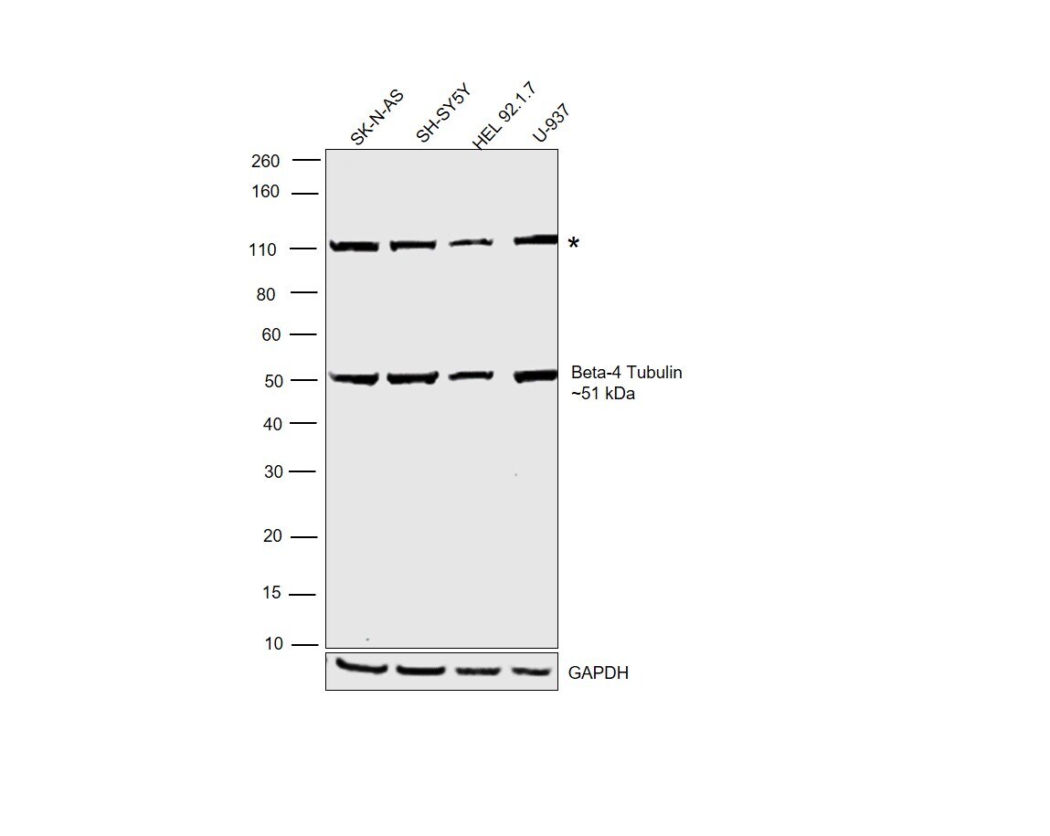
- Western blot was performed using Anti-beta-4 Tubulin Polyclonal Antibody (Product # PA5-21416) and a 51 kDa band corresponding to beta-4 Tubulin was observed across the cell lines tested. A non-specific band (*) was also observed at ~110 kDa. Whole-cell extracts (30 µg lysate) of SK-N-AS (Lane 1), SH-SY5Y (Lane 2), HEL 92.1.7 (Lane 3), and U-937 (Lane 4) were electrophoresed using NuPAGE™ 4-12% Bis-Tris Protein Gel (Product # NP0322BOX). Resolved proteins were then transferred onto a nitrocellulose membrane (Product # IB23001) by iBlot® 2 Dry Blotting System (Product # IB21001). The blot was probed with the primary antibody (1:1000 dilution) and detected by chemiluminescence with Goat anti-Rabbit IgG (H+L) Superclonal™ Recombinant Secondary Antibody, HRP (Product # A27036,1:20,000 dilution) using the iBright™ FL1500 Imaging System (Product # A44115). Chemiluminescent detection was performed using SuperSignal™ West Pico PLUS Chemiluminescent Substrate (Product # 34580).
- Submitted by
- Invitrogen Antibodies (provider)
- Main image
- Experimental details
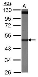
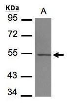
- Western Blot using beta-4 Tubulin Polyclonal Antibody (Product # PA5-21416). Sample (30 µg of whole cell lysate). A: 293T. 7.5% SDS PAGE. Beta-4 Tubulin Polyclonal Antibody (Product # PA5-21416) diluted at 1:1,000. The HRP-conjugated anti-rabbit IgG antibody was used to detect the primary antibody.
Supportive validation
- Submitted by
- Invitrogen Antibodies (provider)
- Main image

- Experimental details
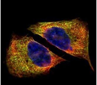
- Immunofluorescent analysis of beta IV Tubulin in paraformaldehyde-fixed U2OS cells using a beta IV Tubulin polyclonal antibody (Product # PA5-21416) (Green) at a 1:500 dilution. Alpha-tubulin filaments were labeled with Product # PA5-29281 (Red) at a 1:2000.
- Submitted by
- Invitrogen Antibodies (provider)
- Main image

- Experimental details
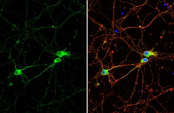
- Immunocytochemistry-Immunofluorescence analysis of beta-4-Tubulin was performed in DIV9 rat E18 primary cortical neuron cells fixed in 4% paraformaldehyde at RT for 15 min. Green: beta-4 Tubulin Polyclonal Antibody (Product # PA5-21416) diluted at 1:500. Red: beta Tubulin 3/ Tuj1, stained by beta Tubulin 3/ Tuj1 antibody. Blue: Fluoroshield with DAPI.
- Submitted by
- Invitrogen Antibodies (provider)
- Main image
- Experimental details
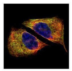
- Confocal immunofluorescence analysis (Olympus FV10i) of paraformaldehyde-fixed U2OS, using beta Tubulin 4 antibody (Product # PA5-21416) (Green) at 1:500 dilution. Alpha-tubulin filaments were labeled with (Product # MA1-25054) (Red) at 1:2,000.

- Submitted by
- Invitrogen Antibodies (provider)
- Main image
- Experimental details
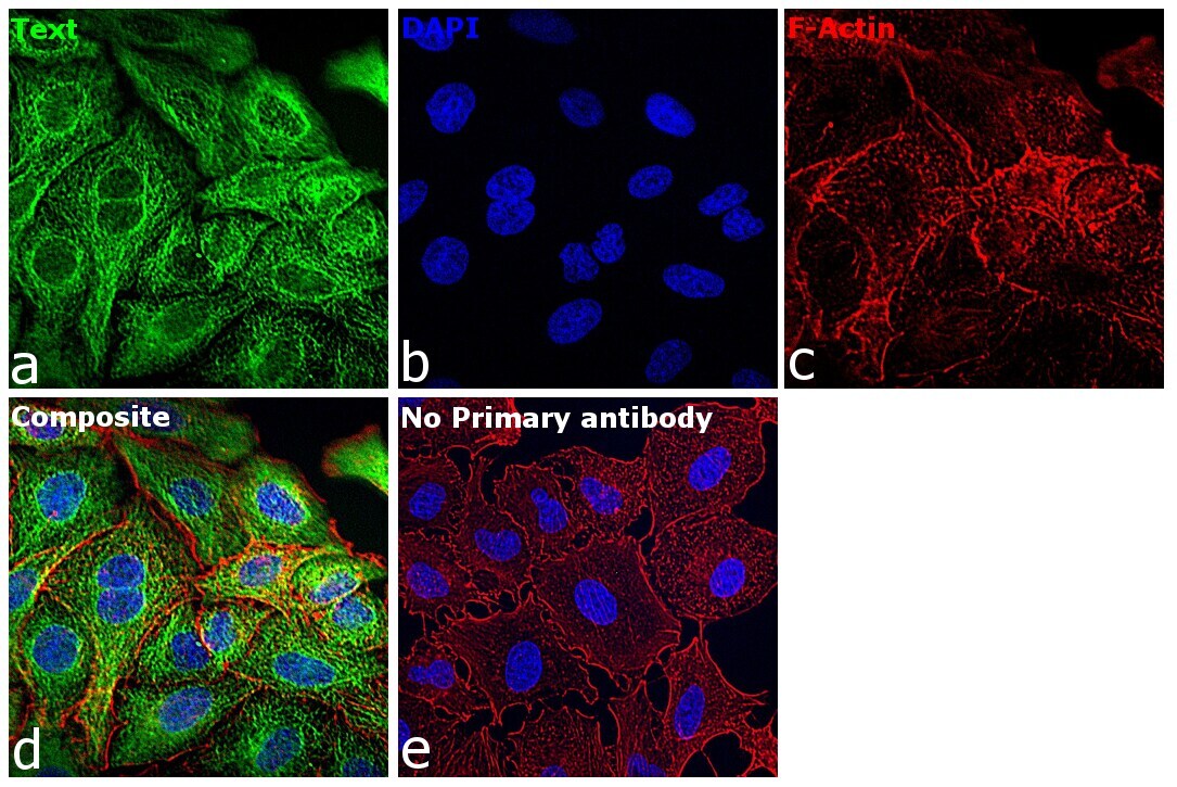
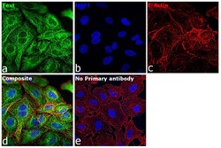
- Immunofluorescence analysis of beta-4 Tubulin was performed using 70 confluent log phase A549 cells. The cells were fixed with 4% paraformaldehyde for 10 minutes, permeabilized with 0.1% Triton™ X-100 for 15 minutes, and blocked with 2% BSA for 45 minutes at room temperature. The cells were labeled with beta-4 Tubulin Polyclonal Antibody (Product # PA5-21416) at 1:100 dilution in 0.1% BSA, incubated at 4 degrees celsius overnight, and then labeled with Donkey anti-Rabbit IgG (H+L) Highly Cross-Adsorbed Secondary Antibody, Alexa Fluor Plus 488 (Product # A32790), (1:2000 dilution), for 45 minutes at room temperature (Panel a: Green). Nuclei (Panel b: Blue) were stained with Hoechst 33342 (Product # H1399). F-actin (Panel c: Red) was stained with Alexa Fluor™ Plus 647 Phalloidin (Product # A30107, 1:2000 dilution). Panel d represents the merged image showing cytoskeletal localization. Panel e represents control cells with no primary antibody to assess the background. The images were captured at 40X magnification in CellInsight CX7 LZR High-Content Screening (HCS) Platform (Product # CX7A1110LZR) and externally deconvoluted (D.Sage et al. / Methods 115 (2017) 28–41).