Explore
 Validate
 Learn
Learn Western blot
 Immunohistochemistry
Antibody data
- Antibody Data
- Antigen structure
- References [1]
- Comments [0]
- Validations
- Immunohistochemistry [1]
Submit
Validation data
Reference
Comment
Report error
- Product number
- AF2459 - Provider product page

- Provider
- Novus Biologicals
- Product name
- Goat Polyclonal RGM-A Antibody
- Antibody type
- Polyclonal
- Description
- Antigen Affinity-purified. Detects human RGM-A in direct ELISAs and Western blots. In direct ELISAs, less than 40% cross-reactivity with recombinant chicken RGM and recombinant mouse RGM-A is observed, and less than 1% cross-reactivity with recombinant human (rh) RGM-B and rhRGM-C is observed..
- Reactivity
- Human
- Host
- Goat
- Conjugate
- Unconjugated
- Isotype
- IgG
- Vial size
- 100 ug
- Concentration
- LYOPH
- Storage
- Use a manual defrost freezer and avoid repeated freeze-thaw cycles. 12 months from date of receipt, -20 to -70 degreesC as supplied. 1 month, 2 to 8 degreesC under sterile conditions after reconstitution. 6 months, -20 to -70 degreesC under sterile conditions after reconstitution.
Submitted references The Netrin-4/ Neogenin-1 axis promotes neuroblastoma cell survival and migration.
Villanueva AA, Falcón P, Espinoza N, R LS, Milla LA, Hernandez-SanMiguel E, Torres VA, Sanchez-Gomez P, Palma V
Oncotarget 2017 Feb 7;8(6):9767-9782
Oncotarget 2017 Feb 7;8(6):9767-9782
No comments: Submit comment
Supportive validation
- Submitted by
- Novus Biologicals (provider)
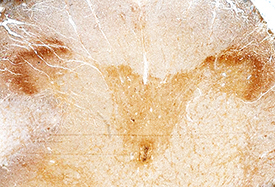
- Main image

- Experimental details
- RGM-A in Human Brain. RGM-A was detected in immersion fixed paraffin-embedded sections of human brain (spinal cord) using Goat Anti-Human RGM-A Antigen Affinity-purified Polyclonal Antibody (Catalog # AF2459) at 15 µg/mL overnight at 4 °C. Before incubation with the primary antibody, tissue was subjected to heat-induced epitope retrieval using Antigen Retrieval Reagent-Basic (Catalog # CTS013). Tissue was stained using the Anti-Goat HRP-DAB Cell & Tissue Staining Kit (brown; Catalog # CTS008) and counterstained with hematoxylin (blue). Specific staining was localized to the dorsal horn. View our protocol for Chromogenic IHC Staining of Paraffin-embedded Tissue Sections.