 Explore
 Validate
 Learn
Learn Western blot
 Immunocytochemistry
Antibody data
- Antibody Data
- Antigen structure
- References [0]
- Comments [0]
- Validations
- Immunocytochemistry [3]
- Immunoprecipitation [1]
- Immunohistochemistry [4]
- Other assay [1]
Submit
Validation data
Reference
Comment
Report error
- Product number
- PA5-63558 - Provider product page

- Provider
- Invitrogen Antibodies
- Product name
- TMEM106B Polyclonal Antibody
- Antibody type
- Polyclonal
- Antigen
- Recombinant protein fragment
- Description
- Immunogen sequence: GKSLSHLPLH SSKEDAYDGV TSENMRNGLV NSEVHNEDGR NGDVSQFPYV EF Highest antigen sequence identity to the following orthologs: Mouse - 91%, Rat - 87%.
- Reactivity
- Human
- Host
- Rabbit
- Isotype
- IgG
- Vial size
- 100 μL
- Concentration
- 0.1 mg/mL
- Storage
- Store at 4°C short term. For long term storage, store at -20°C, avoiding freeze/thaw cycles.
No comments: Submit comment
Supportive validation
- Submitted by
- Invitrogen Antibodies (provider)
- Main image
- Experimental details
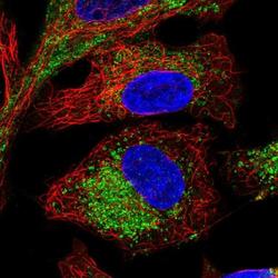
- Immunofluorescent staining of TMEM106B in human cell line A549 shows positivity in vesicles. Samples were probed using a TMEM106B Polyclonal Antibody (Product # PA5-63558).
- Submitted by
- Invitrogen Antibodies (provider)
- Main image
- Experimental details
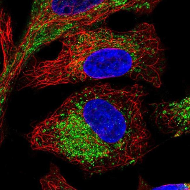
- Immunofluorescent staining of TMEM106B in human cell line A549 using a TMEM106B Polyclonal Antibody (Product # PA5-63558) shows localization to endosomes and lysosomes.
- Submitted by
- Invitrogen Antibodies (provider)
- Main image

- Experimental details
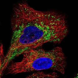
- Immunofluorescent staining of TMEM106B in human cell line A549 using a TMEM106B Polyclonal Antibody (Product # PA5-63558) shows localization to endosomes and lysosomes.

Supportive validation
- Submitted by
- Invitrogen Antibodies (provider)
- Main image

- Experimental details
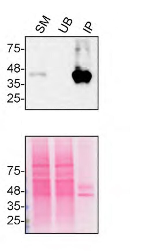
- Immunoprecipitation of TMEM106B was performed on HAP1 WT cell lysate. Antibody-bead conjugate was prepared by adding 2 µg of TMEM106B Polyclonal Antibody (Product # PA5-63558) with 30 µL of Dynabeads™ Protein A (Product # 10002D) and rocked for ~2 hour at 4 degree celcius. One mg of protein was incubated with the antibody-bead conjugate for ~2 hours at 4 degree celcius. Following centrifugation and multiple washes, 2% starting material (SM), 2% unbound fraction (UB) and immunoprecipitated fraction (IP) were processed for immunoblot using a different antibody. Ponceau stained transfer of blot is shown (below immunoblot). Data courtesy of YCharOS Inc., an open science company with the mission of characterizing commercially available antibodies using knockout validation.
Supportive validation
- Submitted by
- Invitrogen Antibodies (provider)
- Main image
- Experimental details
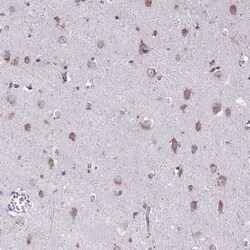
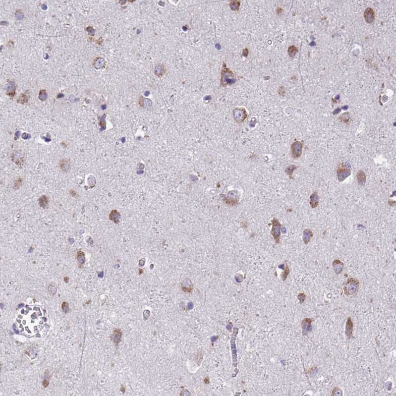
- Immunohistochemical staining of TMEM106B in human cerebral cortex using TMEM106B Polyclonal Antibody (Product # PA5-63558) shows positivity in neuronal cells.
- Submitted by
- Invitrogen Antibodies (provider)
- Main image
- Experimental details
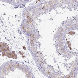
- Immunohistochemical staining of TMEM106B in human testis using TMEM106B Polyclonal Antibody (Product # PA5-63558) shows cytoplasmic positivity in cells in seminiferous ducts and in Leydig cells..

- Submitted by
- Invitrogen Antibodies (provider)
- Main image

- Experimental details
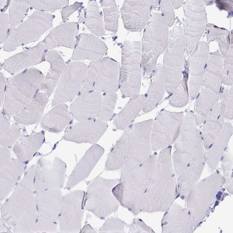
- Immunohistochemical staining of TMEM106B in human skeletal muscle using TMEM106B Polyclonal Antibody (Product # PA5-63558) shows no positivity in myocytes as expected.
- Submitted by
- Invitrogen Antibodies (provider)
- Main image
- Experimental details
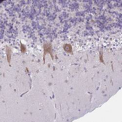
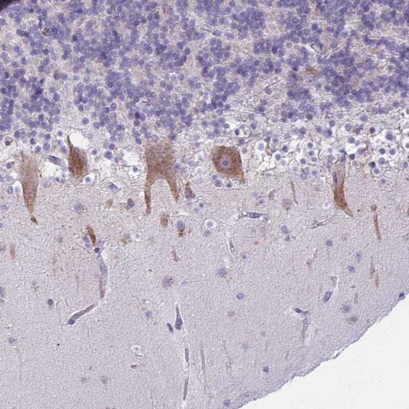
- Immunohistochemical staining of TMEM106B in human cerebellum using TMEM106B Polyclonal Antibody (Product # PA5-63558) shows positivity in Purkinje cells.
Supportive validation
- Submitted by
- Invitrogen Antibodies (provider)
- Main image

- Experimental details
- Immunoprecipitation of TMEM106B was performed on HAP1 WT cell lysate. Antibody-bead conjugate was prepared by adding 2 µg of TMEM106B Polyclonal Antibody (Product # PA5-63558) with 30 µL of Dynabeads™ Protein A (Product # 10002D) and rocked for ~2 hour at 4 degree celcius. One mg of protein was incubated with the antibody-bead conjugate for ~2 hours at 4 degree celcius. Following centrifugation and multiple washes, 2% starting material (SM), 2% unbound fraction (UB) and immunoprecipitated fraction (IP) were processed for immunoblot using a different antibody. Ponceau stained transfer of blot is shown (below immunoblot). Data courtesy of YCharOS Inc., an open science company with the mission of characterizing commercially available antibodies using knockout validation.
