 Explore
 Validate
 Learn
LearnAP7637a
antibody from Abcepta
Targeting: FGFR2
BEK, CD332, CEK3, CFD1, ECT1, JWS, K-SAM, KGFR, TK14, TK25
 Western blot
Antibody data
- Antibody Data
- Antigen structure
- References [2]
- Comments [0]
- Validations
- Western blot [1]
- Immunocytochemistry [2]
- Immunohistochemistry [1]
- Flow cytometry [1]
Submit
Validation data
Reference
Comment
Report error
- Product number
- AP7637a - Provider product page

- Provider
- Abcepta
- Proper citation
- Abgent Cat#AP7637a, RRID:AB_2278342
- Product name
- FGFR2 Antibody (N-term)
- Antibody type
- Polyclonal
- Antigen
- Synthetic peptide
- Description
- Peptide Affinity Purified Rabbit Polyclonal Antibody (Pab)
- Reactivity
- Human, Mouse, Rat, Simian
- Host
- Rabbit
- Isotype
- IgG
- Vial size
- 400 µl
- Concentration
- 0.5 mg/ml
- Storage
- Maintain refrigerated at 2-8°C for up to 6 months. For long term storage store at -20°C in small aliquots to prevent freeze-thaw cycles.
Submitted references Induction of stem cell gene expression in adult human fibroblasts without transgenes.
E2F-1-deficient NOD/SCID mice developed showing decreased saliva production.
Page RL, Ambady S, Holmes WF, Vilner L, Kole D, Kashpur O, Huntress V, Vojtic I, Whitton H, Dominko T
Cloning and stem cells 2009 Sep;11(3):417-26
Cloning and stem cells 2009 Sep;11(3):417-26
E2F-1-deficient NOD/SCID mice developed showing decreased saliva production.
Matsui-Inohara H, Uematsu H, Narita T, Satoh K, Yonezawa H, Kuroda K, Ito T, Yoneda S, Kawarai T, Sugiya H, Watanabe H, Senpuku H
Experimental biology and medicine (Maywood, N.J.) 2009 Dec;234(12):1525-36
Experimental biology and medicine (Maywood, N.J.) 2009 Dec;234(12):1525-36
No comments: Submit comment
Supportive validation
- Submitted by
- Abcepta (provider)
- Main image

- Experimental details
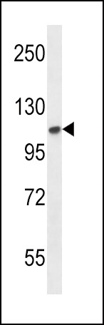
- FGFR2 Antibody (N-term) (Cat. #AP7637a) western blot analysis in mouse NIH-3T3 cell line lysates (35ug/lane).This demonstrates the FGFR2 antibody detected the FGFR2 protein (arrow).
- Primary Ab dilution
- 1:1000
Supportive validation
- Submitted by
- Abcepta (provider)
- Main image
- Experimental details
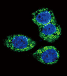
- Confocal immunofluorescent analysis of FGFR2 Antibody (N-term)(Cat#AP7637a) with Hela cell followed by Alexa Fluor 488-conjugated goat anti-rabbit lgG (green).DAPI was used to stain the cell nuclear (blue).
- Primary Ab dilution
- 1:10~50

- Submitted by
- Abcepta (provider)
- Main image

- Experimental details
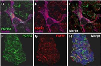
- C:FGFR2/isolectinB4 (C) and FGFR1/isolectinB4 (D) staining of apparent mesenchymal cells and the subpopulation of endothelial cells. Virtually all other dispersed apparent mesenchymal cells express FGFR1 and FGFR2 (merged image in E). F: FGFR2 (F) and FGFR1 (G) staining in clustered cells of epithelial origin (inferred by morphology here) demonstrating that epithelial cells express both FGFR1 and FGFR2 (merged image with DAPI staining in H).
- Primary Ab dilution
- 1:50~100
Supportive validation
- Submitted by
- Abcepta (provider)
- Main image
- Experimental details
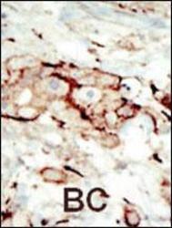
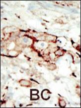
- "Formalin-fixed and paraffin-embedded human cancer tissue reacted with the primary antibody, which was peroxidase-conjugated to the secondary antibody, followed by AEC staining. This data demonstrates the use of this antibody for immunohistochemistry; clinical relevance has not been evaluated. BC = breast carcinoma; HC = hepatocarcinoma."
- Primary Ab dilution
- 1:50~100
Supportive validation
- Submitted by
- Abcepta (provider)
- Main image
- Experimental details
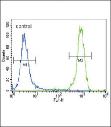
- FGFR2 Antibody (N-term) (Cat. #AP7637a) flow cytometric analysis of NCI-H460 cells (right histogram) compared to a negative control cell (left histogram).FITC-conjugated goat-anti-rabbit secondary antibodies were used for the analysis.
- Primary Ab dilution
- 1:10~50
