Explore
 Validate
 Learn
Learn Western blot
 Immunohistochemistry
Antibody data
- Antibody Data
- Antigen structure
- References [1]
- Comments [0]
- Validations
- Immunohistochemistry [1]
- Other assay [2]
Submit
Validation data
Reference
Comment
Report error
- Product number
- PA5-15000 - Provider product page

- Provider
- Invitrogen Antibodies
- Product name
- ALDH1A3 Polyclonal Antibody
- Antibody type
- Polyclonal
- Antigen
- Synthetic peptide
- Reactivity
- Human, Mouse
- Host
- Rabbit
- Isotype
- IgG
- Vial size
- 400 μL
- Concentration
- 2 mg/mL
- Storage
- Store at 4°C short term. For long term storage, store at -20°C, avoiding freeze/thaw cycles.
Submitted references ALDH1 Bio-activates Nifuroxazide to Eradicate ALDH(High) Melanoma-Initiating Cells.
Sarvi S, Crispin R, Lu Y, Zeng L, Hurley TD, Houston DR, von Kriegsheim A, Chen CH, Mochly-Rosen D, Ranzani M, Mathers ME, Xu X, Xu W, Adams DJ, Carragher NO, Fujita M, Schuchter L, Unciti-Broceta A, Brunton VG, Patton EE
Cell chemical biology 2018 Dec 20;25(12):1456-1469.e6
Cell chemical biology 2018 Dec 20;25(12):1456-1469.e6
No comments: Submit comment
Supportive validation
- Submitted by
- Invitrogen Antibodies (provider)
- Main image
- Experimental details
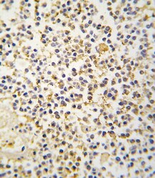
- Immunohistochemistry analysis of ALDH1A3 in formalin-fixed and paraffin-embedded human kidney tissue. Samples were incubated with ALDH1A3 polyclonal antibody (Product # PA5-15000) which was peroxidase-conjugated to the secondary antibody, followed by DAB staining. This data demonstrates the use of this antibody for immunohistochemistry; clinical relevance has not been evaluated.

Supportive validation
- Submitted by
- Invitrogen Antibodies (provider)
- Main image
- Experimental details
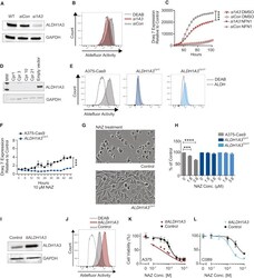
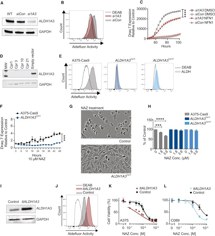
- Figure 3 ALDH1A3 Mediates Nifuroxazide Activity (A) Western blot of ALDH1A3 in ALDH1A3 siRNA transfected A375 ( si1A3 ) cells, siRNA control ( siCon ), and wild-type (WT) control cells. GAPDH, loading control. (B) Aldefluor activity in si1A3 cells or siCon cells. DEAB was used as a negative control (n > 3). (C) Sensitivity of si1A3 and siCon cells to NFN1 measured by levels of Draq7 expression using IncuCyte Zoom. Values are means +- SEM (n = 3, ****p < 0.0001, ANOVA with Tukey's test). (D) Western blot of ALDH1A3 protein in ALDH1A3 knockout single-cell clones. GAPDH, loading control (n > 3). (E) Aldefluor activity in ALDH1A3 knockout clones and A375-Cas9 WT cells. DEAB is used as negative control. (F) Sensitivity of ALDH1A3 Cpr3 and A375-Cas9 cells to 10 muM nifuroxazide measured by levels of Draq7 expression using IncuCyte Zoom. Values are means +- SEM (n = 4, ***p < 0.001, ANOVA). (G) Representative images of WT and ALDH1A3 Cpr3 cells treated with nifuroxazide or DMSO, imaged with IncuCyte Zoom. Note cell death in upper panel. (H) Clonogenic potential of ALDH1A3 Cpr3 , ALDH1A3 Cpr21 , and control A375-Cas9 cells in soft agar. Values are means +- SEM (n = 3, ***p < 0.001, ****p < 0.0001, ANOVA with Dunnett's test). (I) Western blot image of ALDH1A3 protein levels in A375 cells overexpressing ALDH1A3 (transient transfection ALDH1A3 [ ttALDH1A3 ]) and WT control cells. GAPDH, loading control. (J) Aldefluor activity in ttALDH1A3 and WT control. DEAB w
- Submitted by
- Invitrogen Antibodies (provider)
- Main image
- Experimental details
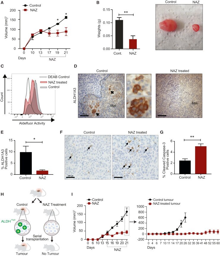
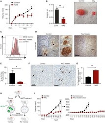
- Figure 5 Nifuroxazide Targets ALDH1A3 High Subpopulations in Melanoma Tumors (A) Sensitivity of A375-L2T tumors to nifuroxazide in vivo . Mice were treated with 150 mg/kg nifuroxazide or vehicle for 9 continuous days. Values are means +- SEM (n = 3 mice/condition; experimental replicates >3, *p < 0.05, two-way ANOVA with Sidak's test). (B) Tumor weights of control or nifuroxazide-treated mice at the end of drug treatment. Values are means +- SEM (n = 3 mice/condition; experimental replicates >3, **p < 0.01; Student's t test). Image of tumors from control or nifuroxazide-treated mice at the end of drug treatment duration. (C) Aldefluor activity in A375-L2T cells in tumors treated with nifuroxazide or vehicle for 9 days. Note the significant shift to the left in the tumors from nifuroxazide-treated mice indicating reduced Aldefluor activity (geometric mean of Aldefluor activity; n = 3 p < 0.05; Student's t test). (D) Immunohistochemistry staining of ALDH1A3 expression in melanoma tumors generated from A375-L2T cells treated with nifuroxazide or vehicle for 5 days (n = 3). 20x. Scale bar, 100 mum. Arrows point to ALDH1 High cell clusters. Mel, melanoma tumor. Dotted lines outline tumor. (E) Quantification of ALDH1A3-positive cells in melanoma tumors treated with nifuroxazide or vehicle. The percentage mean of ALDH1A3-positive cells in melanoma tumors treated with nifuroxazide or vehicle from 10 random fields of view with 10x magnification is presented (*p < 0.05; Student's t