 Explore
 Validate
 Learn
Learn Western blot
Antibody data
- Antibody Data
- Antigen structure
- References [0]
- Comments [0]
- Validations
- Western blot [3]
- Immunocytochemistry [3]
- Immunohistochemistry [1]
Submit
Validation data
Reference
Comment
Report error
- Product number
- PA1-32348 - Provider product page

- Provider
- Invitrogen Antibodies
- Product name
- NEFM Polyclonal Antibody
- Antibody type
- Polyclonal
- Antigen
- Recombinant full-length protein
- Description
- PA1-32348 detects NEFM from rat samples.
- Reactivity
- Human, Mouse, Rat, Bovine, Chicken/Avian, Feline, Porcine
- Host
- Rabbit
- Isotype
- IgG
- Vial size
- 50 µL
- Concentration
- Conc. Not Determined
- Storage
- Store at 4°C short term. For long term storage, store at -20°C, avoiding freeze/thaw cycles.
No comments: Submit comment
Supportive validation
- Submitted by
- Invitrogen Antibodies (provider)
- Main image

- Experimental details
- Western Blot analysis of rat cerebellum tissue using NEFM Polyclonal Antibody (Product # PA1-32348).

- Submitted by
- Invitrogen Antibodies (provider)
- Main image

- Experimental details
- Western blot analysis of NEFM was performed by loading 20 µg of HEK-293 wild type (Lane 1), HEK-293 Cas9 control (Lane 2), HEK-293 NEFM knockout (Lane 3) membrane enriched cell extracts. The blot was probed with Anti-NEFM Polyclonal Antibody (Product # PA1-32348) (1:2000 dilution) and Goat anti-Rabbit IgG (H+L), Superclonal™ Recombinant Secondary Antibody, HRP (Product # A27036) (1:4000 dilution). Loss of signal upon CRISPR mediated knockout (KO) confirms that antibody is specific to NEFM.

- Submitted by
- Invitrogen Antibodies (provider)
- Main image

- Experimental details
- Western blot was performed using Anti-NEFM Polyclonal Antibody (Product # PA1-32348) and a 160 kDa band corresponding to NEFM was observed in HEK-293 cell line, Mouse Cerebellum, Rat Brain but not in HeLa, Hep G2 cell line, Mouse Heart and Mouse Spleen. Membrane extracts (30 µg lysate) of HEK-293 (Lane 1), HeLa (Lane 2) and Hep G2 (Lane 3), Mouse Cerebellum (Lane 4), Mouse Brain (Lane 5), Rat Brain (Lane 6), Mouse Heart (Lane 7) and Mouse Spleen (Lane 8) were electrophoresed using Novex® NuPAGE® 4-12 % Bis-Tris gel (Product # NP0321BOX). Resolved proteins were then transferred onto a nitrocellulose membrane (Product # IB23001) by iBlot® 2 Dry Blotting System (Product # IB21001). The blot was probed with the primary antibody (1:5000 dilution) and detected by chemiluminescence with Goat anti-Rabbit IgG (H+L), Superclonal™ Recombinant Secondary Antibody, HRP (Product # A27036, 1:4000 dilution) using the iBright FL 1000 (Product # A32752). Chemiluminescent detection was performed using Novex® ECL Chemiluminescent Substrate Reagent Kit (Product # WP20005).

Supportive validation
- Submitted by
- Invitrogen Antibodies (provider)
- Main image
- Experimental details
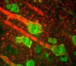
- Immunofluorescent analysis of NEFM in sections of rat cerebral cortex using a NEFM polyclonal antibody (Product # PA1-32348) (red), which reveals the perikarya of pyramidal neurons and dendrites and axons surrounding them. The green channel shows staining with a monoclonal antibody to the beta-adrendergic receptor kinase 1.

- Submitted by
- Invitrogen Antibodies (provider)
- Main image
- Experimental details
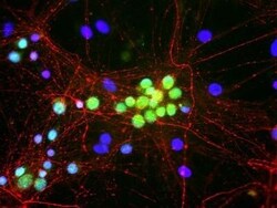
- Immunocytochemistry-Immunofluorescence analysis of NEFM in mixed neuron/glia cell cultures using NEFM Polyclonal Antibody (Product # PA1-32348) (Red). Green: Fox1 antibody . Blue: DAPI.

- Submitted by
- Invitrogen Antibodies (provider)
- Main image
- Experimental details
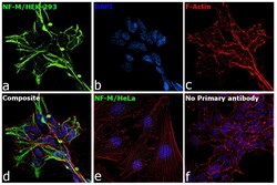
- Immunofluorescence analysis of NF-M was performed using 70% confluent log phase HEK-293 and HeLa cells. The cells were fixed with 4% paraformaldehyde for 10 minutes, permeabilized with 0.1% Triton™ X-100 for 15 minutes, and blocked with 2% BSA for 1 hour at room temperature. HEK-293 cells were labeled with NF-M Rabbit Polyclonal Antibody (Product # PA1-32348) at 1:250 dilution in 0.1% BSA, incubated at 4 degree Celsius overnight and then labeled with Goat anti-Rabbit IgG (H+L) Superclonal™ Recombinant Secondary Antibody, Alexa Fluor® 488 conjugate (Product # A27034) at a dilution of 1:2000 for 45 minutes at room temperature (Panel a: green). Nuclei (Panel b: blue) were stained with ProLong™ Diamond Antifade Mountant with DAPI (Product # P36962). F-actin (Panel c: red) was stained with Rhodamine Phalloidin (Product # R415). Panel d represents the merged image of HEK-293 showing cytoskeletal (intermediate filaments) localization. Panel e represents the merged image of HeLa cells showing no expression for NF-M protein. Panel f represents control cells with no primary antibody to assess background. The images were captured at 60X magnification.

Supportive validation
- Submitted by
- Invitrogen Antibodies (provider)
- Main image

- Experimental details
- Immunohistochemistry (Frozen) analysis of NEFM was performed in rat cerebral cortex tissue. Red: NEFM Polyclonal Antibody (Product # PA1-32348) diluted at 1:5000. Green: beta-adrendergic receptor kinase 1. This immunostaining reveals the perikarya of pyramidal neurons and dendrites as well as axons surrounding the neurons.
