Explore
 Validate
 Learn
Learn Western blot
 ELISA
ELISA Immunocytochemistry
 Immunohistochemistry
Antibody data
- Antibody Data
- Antigen structure
- References [0]
- Comments [0]
- Validations
- Western blot [1]
- Immunohistochemistry [1]
Submit
Validation data
Reference
Comment
Report error
- Product number
- LS-C354909 - Provider product page

- Provider
- LSBio
- Product name
- NEFM / NF-M Antibody (clone RMO 14.9) LS-C354909
- Antibody type
- Monoclonal
- Description
- Protein A purified
- Reactivity
- Human, Mouse, Rat, Chicken/Avian, Rabbit
- Host
- Mouse
- Isotype
- IgG
- Antibody clone number
- RMO 14.9
- Storage
- Store at -20°C. Avoid freeze-thaw cycles.
No comments: Submit comment
Enhanced validation
- Submitted by
- LSBio (provider)
- Enhanced method
- Genetic validation
- Main image
- Experimental details
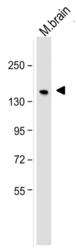
- Western blot analysis of neurofilament, medium chain was performed by loading 25ug of mouse brain tissue lysate per well onto a polyacrylamide gel. Proteins were transferred to a PVDF membrane and blocked. NF-M was detected at ~160kD using a neurofilament, medium chain antibody at a dilution of 2 µg/mL in blocking buffer overnight at 4C, followed by a HRP-labeled secondary antibody for 1 hour at room temperature and detection with a chemiluminescent substrate.

Supportive validation
- Submitted by
- LSBio (provider)
- Enhanced method
- Genetic validation
- Main image
- Experimental details
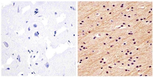
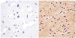
- Immunohistochemistry analysis of the neurofilament medium chain showing staining in the filaments of paraffin-embedded human brain tissue (right) compared to a negative control without primary antibody (left). To expose target proteins, antigen retrieval was performed using 10mM sodium citrate (pH 6.0) and microwaved for 8-15 min. Following antigen retrieval, tissues were blocked in 3% H2O2-methanol for 15 min at room temperature, washed with ddH2O and PBS, and then probed with a Neurofilament medium chain monoclonal antibody diluted in 3% BSA-PBS at a dilution of 1:20 overnight at 4°C in a humidified chamber. Tissues were washed extensively in PBST and detection was performed using an HRP-conjugated secondary antibody followed by colorimetric detection using a DAB kit. Tissues were counterstained with hematoxylin and dehydrated with ethanol and xylene to prep for mounting.