Explore
 Validate
 Learn
Learn Western blot
 Immunocytochemistry
Antibody data
- Antibody Data
- Antigen structure
- References [2]
- Comments [0]
- Validations
- Western blot [2]
- Other assay [1]
Submit
Validation data
Reference
Comment
Report error
- Product number
- NB300-134 - Provider product page

- Provider
- Novus Biologicals
- Proper citation
- Novus Cat#NB300-134, RRID:AB_10000761
- Product name
- Mouse Monoclonal NF-M Antibody
- Antibody type
- Monoclonal
- Description
- Unpurified. Specifically recognizes the medium neurofilament subunit (~145-160 kDa).
- Reactivity
- Human, Mouse, Rat, Bovine, Chicken/Avian, Porcine
- Host
- Mouse
- Isotype
- IgG
- Vial size
- 0.05 ml
- Storage
- Store at 4C short term. Aliquot and store at -20C long term. Avoid freeze-thaw cycles.
Submitted references Activation of the Large-Conductance, Voltage, and Ca(2+)- Activated K(+) (BK) Channel in Acute Spinal Cord Injury in the Wistar Rat Is Neuroprotective.
X-linked Christianson syndrome: heterozygous female Slc9a6 knockout mice develop mosaic neuropathological changes and related behavioral abnormalities.
Jacobsen M, Lett K, Barden JM, Simpson GL, Buttigieg J
Frontiers in neurology 2018;9:1107
Frontiers in neurology 2018;9:1107
X-linked Christianson syndrome: heterozygous female Slc9a6 knockout mice develop mosaic neuropathological changes and related behavioral abnormalities.
Sikora J, Leddy J, Gulinello M, Walkley SU
Disease models & mechanisms 2016 Jan;9(1):13-23
Disease models & mechanisms 2016 Jan;9(1):13-23
No comments: Submit comment
Supportive validation
- Submitted by
- Novus Biologicals (provider)
- Main image
- Experimental details
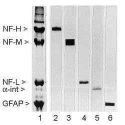
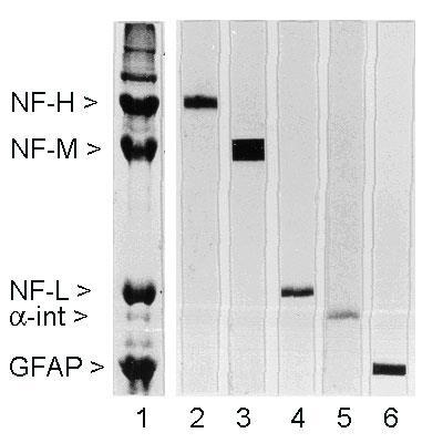
- Western Blot: 160kDa Neurofilament Medium Antibody (3H11) [NB300-134] - Rat spinal cord homogenate showing the major intermediate filament proteins of the nervous system (lane 1). The remaining lanes show blots of this material stainted with various antibodies including NB300-134 (lane 3).
- Submitted by
- Novus Biologicals (provider)
- Main image
- Experimental details
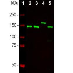
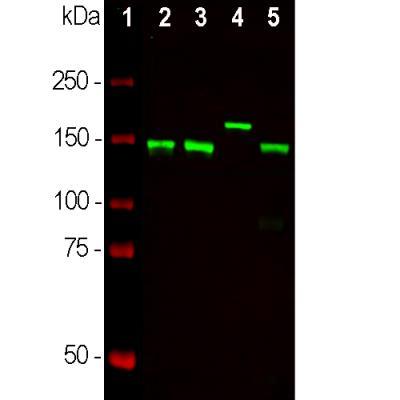
- Western Blot: NF-M Antibody (3H11) [NB300-134] - Neuronal tissue lysates using mouse mAb to NF-M, dilution 1:10,000 in green: [1] protein standard (red), [2] rat spinal cord, [3] mouse spinal cord, [4] cow spinal cord, [5] rat sciatic nerve. Strong bands at ~145 kDa corresponds to rodent NF-M while that at about 160 kDa corresponds to the significantly larger bovine NF-M protein.
Supportive validation
- Submitted by
- Novus Biologicals (provider)
- Main image
- Experimental details
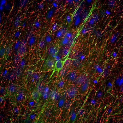
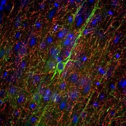
- Immunohistochemistry Free-Floating: NF-M Antibody (3H11) [NB300-134] - Adult rat frontal cortex section stained with mouse mAb to neurofilament NF-M, dilution 1:5,000 in green, and costained with chicken pAb to neurofilament NF-H, dilution 1:5,000 in red. Following transcardial perfusion of rat with 4% paraformaldehyde, brain was post fixed for 24 hours, cut to 45uM, and free-floating sections were stained with above antibodies. NB300-134 antibody labels neuron cell bodies and dendrites of pyramidal neurons, as well as dendrites and axons of other neuronal cells, while the NF-H antibody stains the network of neuronal axons only.