Explore
 Validate
 Learn
Learn Western blot
 Immunocytochemistry
Antibody data
- Antibody Data
- Antigen structure
- References [6]
- Comments [0]
- Validations
- Western blot [1]
- Other assay [1]
Submit
Validation data
Reference
Comment
Report error
- Product number
- NB300-222 - Provider product page

- Provider
- Novus Biologicals
- Proper citation
- Novus Cat#NB300-222, RRID:AB_10002431
- Product name
- Chicken Polyclonal NF-M Antibody
- Antibody type
- Polyclonal
- Description
- Ammonium sulfate precipitation.
- Reactivity
- Human, Mouse, Rat, Bovine, Chicken/Avian, Drosophila, Porcine
- Host
- Chicken/Avian
- Isotype
- IgY
- Vial size
- 0.25 ml
- Storage
- Store at 4C short term. Aliquot and store at -20C long term. Avoid freeze-thaw cycles.
Submitted references Muscleblind acts as a modifier of FUS toxicity by modulating stress granule dynamics and SMN localization.
Axonal and myelinic pathology in 5xFAD Alzheimer's mouse spinal cord.
Tumor suppressor menin is required for subunit-specific nAChR α5 transcription and nAChR-dependent presynaptic facilitation in cultured mouse hippocampal neurons.
The mitochondrial division inhibitor Mdivi-1 rescues mammalian neurons from anesthetic-induced cytotoxicity.
Mechanisms of action of naturally occurring antibodies against β-amyloid on microglia.
Turning placenta into brain: placental mesenchymal stem cells differentiate into neurons and oligodendrocytes.
Casci I, Krishnamurthy K, Kour S, Tripathy V, Ramesh N, Anderson EN, Marrone L, Grant RA, Oliver S, Gochenaur L, Patel K, Sterneckert J, Gleixner AM, Donnelly CJ, Ruepp MD, Sini AM, Zuccaro E, Pennuto M, Pasinelli P, Pandey UB
Nature communications 2019 Dec 6;10(1):5583
Nature communications 2019 Dec 6;10(1):5583
Axonal and myelinic pathology in 5xFAD Alzheimer's mouse spinal cord.
Chu TH, Cummins K, Sparling JS, Tsutsui S, Brideau C, Nilsson KPR, Joseph JT, Stys PK
PloS one 2017;12(11):e0188218
PloS one 2017;12(11):e0188218
Tumor suppressor menin is required for subunit-specific nAChR α5 transcription and nAChR-dependent presynaptic facilitation in cultured mouse hippocampal neurons.
Getz AM, Xu F, Visser F, Persson R, Syed NI
Scientific reports 2017 May 11;7(1):1768
Scientific reports 2017 May 11;7(1):1768
The mitochondrial division inhibitor Mdivi-1 rescues mammalian neurons from anesthetic-induced cytotoxicity.
Xu F, Armstrong R, Urrego D, Qazzaz M, Pehar M, Armstrong JN, Shutt T, Syed N
Molecular brain 2016 Mar 24;9:35
Molecular brain 2016 Mar 24;9:35
Mechanisms of action of naturally occurring antibodies against β-amyloid on microglia.
Gold M, Mengel D, Röskam S, Dodel R, Bach JP
Journal of neuroinflammation 2013 Jan 14;10:5
Journal of neuroinflammation 2013 Jan 14;10:5
Turning placenta into brain: placental mesenchymal stem cells differentiate into neurons and oligodendrocytes.
Portmann-Lanz CB, Schoeberlein A, Portmann R, Mohr S, Rollini P, Sager R, Surbek DV
American journal of obstetrics and gynecology 2010 Mar;202(3):294.e1-294.e11
American journal of obstetrics and gynecology 2010 Mar;202(3):294.e1-294.e11
No comments: Submit comment
Supportive validation
- Submitted by
- Novus Biologicals (provider)
- Main image
- Experimental details
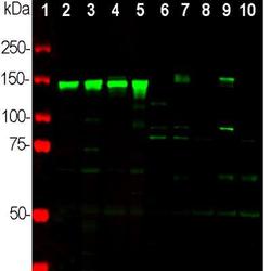
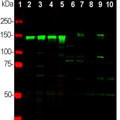
- Western Blot: NF-M Antibody [NB300-222] - Western blot analysis of different neuronal tissue and cell lysates using chicken pAb to NF-M, NB300-222, dilution 1:2,000 in green: [1] protein standard (red), [2] rat brain [3] rat spinal cord, [4] mouse brain, [5] mouse spinal cord, [6] NIH/3T3 cells, [7] HEK293, [8] HeLa, [9] SH-SY5Y, and [10] C6 cells. Strong band at 145kDa corresponds to rodent NF-M, and about 160kDa band corresponds to human NF-M protein, visible in SHSY-5Y and HEK293 cells which have neuronal properties. NF-M is not expressed in HeLa and other cell lines tested.
Supportive validation
- Submitted by
- Novus Biologicals (provider)
- Main image
- Experimental details
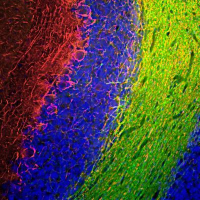
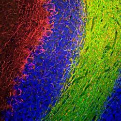
- Immunohistochemistry Free-Floating: NF-M Antibody [NB300-222] - Immunofluorescent analysis of rat cerebellum section stained with chicken pAb to NF-M, NB300-222, dilution 1:1,000 in red, and costained with mouse mAb to CNPase, dilution 1:500, in green. The blue is DAPI staining of nuclear DNA. Following transcardial perfusion of rat with 4% paraformaldehyde, brain was post fixed for 24 hours, cut to 45uM, and free floating sections were stained with above antibodies. The NF-M antibody labels the axons of basket cells and other neurons, while the CNP antibody stains oligodendrocytes, cells that form the myelin sheathes around axons.