Explore
 Validate
 Learn
Learn Western blot
 Immunoprecipitation
Antibody data
- Antibody Data
- Antigen structure
- References [2]
- Comments [0]
- Validations
- Western blot [2]
- Immunocytochemistry [2]
- Immunohistochemistry [4]
Submit
Validation data
Reference
Comment
Report error
- Product number
- MA1-2011 - Provider product page

- Provider
- Invitrogen Antibodies
- Product name
- NEFM Monoclonal Antibody (3H11)
- Antibody type
- Monoclonal
- Antigen
- Synthetic peptide
- Description
- MA1-2011 detects the neurofilament, medium chain in human and rat samples. MA1-2011 has been successfully used in Western blot, immunofluorescent, and immunoprecipitation procedures. By Western blot this antibody detects a ~160 kDa protein representing the neurofilament, medium chain in HEK 293 cells, rat spinal cord, and human frontal cortex. In immunofluorecence procedures MA1-2011 recognizes the neurofilament, medium chain in human neurons. The MA1-2011 immunogen is a synthetic peptide corresponding to a C-terminal fragment of rat neurofilament.
- Reactivity
- Human, Rat
- Host
- Mouse
- Antibody clone number
- 3H11
- Vial size
- 100 µg
- Concentration
- 1 mg/mL
- Storage
- -20° C, Avoid Freeze/Thaw Cycles
Submitted references Preferential transformation of human neuronal cells by human adenoviruses and the origin of HEK 293 cells.
Compartmentation of alpha-internexin and neurofilament triplet proteins in cultured hippocampal neurons.
Shaw G, Morse S, Ararat M, Graham FL
FASEB journal : official publication of the Federation of American Societies for Experimental Biology 2002 Jun;16(8):869-71
FASEB journal : official publication of the Federation of American Societies for Experimental Biology 2002 Jun;16(8):869-71
Compartmentation of alpha-internexin and neurofilament triplet proteins in cultured hippocampal neurons.
Benson DL, Mandell JW, Shaw G, Banker G
Journal of neurocytology 1996 Mar;25(3):181-96
Journal of neurocytology 1996 Mar;25(3):181-96
No comments: Submit comment
Supportive validation
- Submitted by
- Invitrogen Antibodies (provider)
- Main image
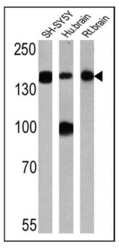
- Experimental details
- Western blot analysis of Neurofilament, Medium chain was performed by loading 25 µg of SH-SY5Y (lane 1), human brain (lane 2) and rat brain (lane 3) lysates onto an SDS polyacrylamide gel. Proteins were transferred to a PVDF membrane and blocked at 4ºC overnight. The membrane was probed with a Neurofilament, Medium chain monoclonal antibody (Product # MA1-2011) at a dilution of 1:500 overnight at 4°C, washed in TBST, and probed with an HRP-conjugated secondary antibody for 1 hr at room temperature in the dark. Chemiluminescent detection was performed using Pierce ECL Plus Western Blotting Substrate (Product # 32132). Results show a band at ~160 kDa.

- Submitted by
- Invitrogen Antibodies (provider)
- Main image
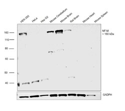
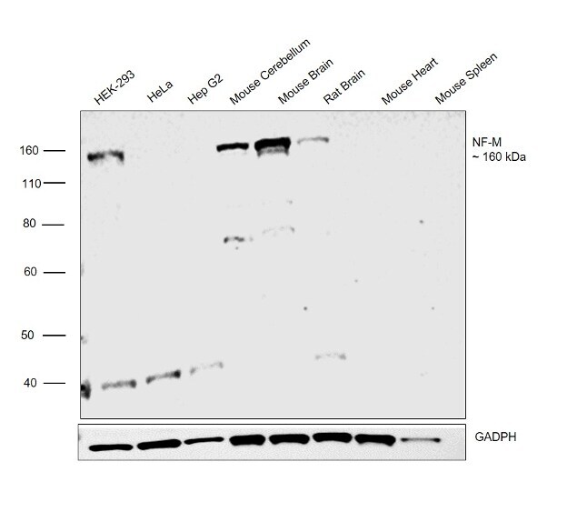
- Experimental details
- Western blot was performed using Anti-NEFM Monoclonal Antibody (3H11) (Product # MA1-2011) and a 160 kDa band corresponding to NEFM was observed in HEK-293 cell line, Mouse Brain, Mouse Cerebellum, Rat Brain but not in HeLa, Hep G2 cell line, Mouse Heart and Mouse Spleen. Membrane extracts (30 µg lysate) of HEK-293 (Lane 1), HeLa (Lane 2) and Hep G2 (Lane 3), Mouse Cerebellum (Lane 4), Mouse Brain (Lane 5), Rat Brain (Lane 6), Mouse Heart (Lane 7) and Mouse Spleen (Lane 8) were electrophoresed using Novex® NuPAGE® 4-12 % Bis-Tris gel (Product # NP0321BOX). Resolved proteins were then transferred onto a nitrocellulose membrane (Product # IB23001) by iBlot® 2 Dry Blotting System (Product # IB21001). The blot was probed with the primary antibody (1:500 dilution) and detected by chemiluminescence with Goat anti-Mouse IgG (H+L), Superclonal™ Recombinant Secondary Antibody, HRP (Product # A28177, 1:4000 dilution) using the iBright FL 1000 (Product # A32752). Chemiluminescent detection was performed using Novex® ECL Chemiluminescent Substrate Reagent Kit (Product # WP20005).
Supportive validation
- Submitted by
- Invitrogen Antibodies (provider)
- Main image

- Experimental details
- Knockout of NEFM was achieved by CRISPR-Cas9 genome editing. Immunofluorescence analysis was performed on wild type HEK-293 cells (panel a,d), HEK-293 Cas9 cells (panels b,e) and HEK-293 NEFM KO cells (panel c,f). Cells were fixed, permeabilized, and labelled with NEFM Monoclonal Antibody(3H11) (Product # MA1-2011) (5 µg/mL), followed by Goat anti-Mouse IgG (H+L) Highly Cross-Adsorbed Secondary Antibody, Alexa Fluor Plus 488 (Product # A32723) (1:2000). Nuclei (blue) were stained using ProLong™ Diamond Antifade Mountant with DAPI (Product # P36962), and Rhodamine Phalloidin (Product # R415) (1:300) was used for cytoskeletal F-actin (red) staining. Loss of signal (panel c,f) upon CRISPR mediated knockout (KO) confirms that antibody is specific to NEFM (green). The images were captured at 60X magnification.

- Submitted by
- Invitrogen Antibodies (provider)
- Main image

- Experimental details
- Immunofluorescence analysis of NF-M was performed using 70% confluent log phase HEK-293 and HeLa cells. The cells were fixed with 4% paraformaldehyde for 10 minutes, permeabilized with 0.1% Triton™ X-100 for 15 minutes, and blocked with 2% BSA for 1 hour at room temperature. HEK-293 cells were labeled with NF-M Mouse Monoclonal Antibody (Product # MA1-2011) at 5 µg/mL in 0.1% BSA, incubated at 4 degree Celsius overnight and then labeled with Goat anti-Mouse IgG (H+L) Superclonal™ Recombinant Secondary Antibody, Alexa Fluor® 488 conjugate (Product # A28175) at a dilution of 1:2000 for 45 minutes at room temperature (Panel a: green). Nuclei (Panel b: blue) were stained with ProLong™ Diamond Antifade Mountant with DAPI (Product # P36962). F-actin (Panel c: red) was stained with Rhodamine Phalloidin (Product # R415). Panel d represents the merged image of HEK-293, positive for NF-M expression showing cytoskeletal (intermediate filaments) localization. Panel e represents the merged image of HeLa cells, that are null for NF-M protein expression. Panel f represents control cells with no primary antibody to assess background. The images were captured at 60X magnification.

Supportive validation
- Submitted by
- Invitrogen Antibodies (provider)
- Main image

- Experimental details
- Immunohistochemistry analysis of Neurofilament Medium Chain showing staining in the cytoplasm of paraffin-embedded rat brain tissue (right) compared to a negative control without primary antibody (left). To expose target proteins, antigen retrieval was performed using 10mM sodium citrate (pH 6.0), microwaved for 8-15 min. Following antigen retrieval, tissues were blocked in 3% H2O2-methanol for 15 min at room temperature, washed with ddH2O and PBS, and then probed with a Neurofilament Medium Chain monoclonal antibody (Product # MA1-2011) diluted in 3% BSA-PBS at a dilution of 1:200 overnight at 4°C in a humidified chamber. Tissues were washed extensively in PBST and detection was performed using an HRP-conjugated secondary antibody followed by colorimetric detection using a DAB kit. Tissues were counterstained with hematoxylin and dehydrated with ethanol and xylene to prep for mounting.

- Submitted by
- Invitrogen Antibodies (provider)
- Main image

- Experimental details
- Immunohistochemistry analysis of Neurofilament Medium Chain showing staining in the cytoplasm of paraffin-embedded rat cerebellum tissue (right) compared to a negative control without primary antibody (left). To expose target proteins, antigen retrieval was performed using 10mM sodium citrate (pH 6.0), microwaved for 8-15 min. Following antigen retrieval, tissues were blocked in 3% H2O2-methanol for 15 min at room temperature, washed with ddH2O and PBS, and then probed with a Neurofilament Medium Chain monoclonal antibody (Product # MA1-2011) diluted in 3% BSA-PBS at a dilution of 1:200 overnight at 4°C in a humidified chamber. Tissues were washed extensively in PBST and detection was performed using an HRP-conjugated secondary antibody followed by colorimetric detection using a DAB kit. Tissues were counterstained with hematoxylin and dehydrated with ethanol and xylene to prep for mounting.

- Submitted by
- Invitrogen Antibodies (provider)
- Main image
- Experimental details
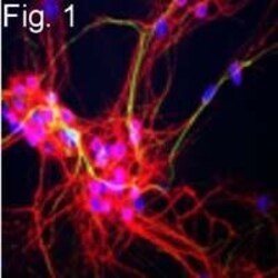
- Figures 1 illustrate immunofluorescence of neurofilament, medium chain in rat cerebral cortex cultures in green using Product # MA1-2011 at 1:200 dilution. MAP2 detection is shown in red, and DAPI is shown in blue.

- Submitted by
- Invitrogen Antibodies (provider)
- Main image

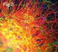
- Experimental details
- Figures 2 illustrate immunofluorescence of neurofilament, medium chain in rat cerebral cortex cultures in green using Product # MA1-2011 at 1:200 dilution. MAP2 detection is shown in red, and DAPI is shown in blue.