Explore
 Validate
 Learn
Learn Western blot
Antibody data
- Antibody Data
- Antigen structure
- References [1]
- Comments [0]
- Validations
- Western blot [3]
- Immunocytochemistry [3]
- Immunohistochemistry [1]
- Other assay [1]
Submit
Validation data
Reference
Comment
Report error
- Product number
- PA1-16758 - Provider product page

- Provider
- Invitrogen Antibodies
- Product name
- NEFM Polyclonal Antibody
- Antibody type
- Polyclonal
- Antigen
- Purifed from natural sources
- Description
- Suggested positive control: antigen standard for NEFM (transient overexpression lysate).
- Reactivity
- Human, Mouse, Rat, Bovine, Chicken/Avian, Drosophila, Porcine
- Host
- Chicken/Avian
- Isotype
- IgY
- Vial size
- 250 µL
- Storage
- Store at 4°C short term. For long term storage, store at -20°C, avoiding freeze/thaw cycles.
Submitted references ROS scavengers decrease γH2ax spots in motor neuronal nuclei of ALS model mice in vitro.
Junghans M, John F, Cihankaya H, Schliebs D, Winklhofer KF, Bader V, Matschke J, Theiss C, Matschke V
Frontiers in cellular neuroscience 2022;16:963169
Frontiers in cellular neuroscience 2022;16:963169
No comments: Submit comment
Supportive validation
- Submitted by
- Invitrogen Antibodies (provider)
- Main image
- Experimental details
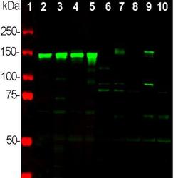
- Western blot analysis of NEFM in neuronal tissue and cell lysates. Samples were incubated in NEFM polyclonal antibody (Product # PA1-16758 using a dilution of 1:2,000. Antibody in green: [1] protein standard (red), [2] rat brain [3] rat spinal cord, [4] mouse brain, [5] mouse spinal cord, [6] NIH/3T3 cells, [7] HEK293, [8] HeLa, [9] SH-SY5Y, and [10] C6 cells. Strong band at 145 kDa corresponds to rodent NF-M, and about 160 kDa band corresponds to human NF-M protein, visible in SHSY-5Y and HEK293 cells which have neuronal properties. NF-M is not expressed in HeLa and other cell lines tested.
- Submitted by
- Invitrogen Antibodies (provider)
- Main image

- Experimental details
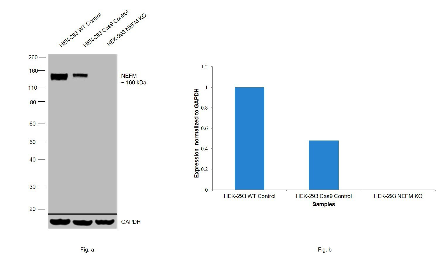
- Western blot analysis of NEFM was performed by loading 20 µg of HEK-293 wild type (Lane 1), HEK-293 Cas9 control (Lane 2), HEK-293 NEFM knockout (Lane 3) membrane enriched cell extracts. The blot was probed with Anti-NEFM Monoclonal Antibody (Product # PA1-16758) (1:5000 dilution) and Goat anti-Chicken IgY (H+L) Secondary Antibody, HRP (Product # A16054). Loss of signal upon CRISPR mediated knockout (KO) confirms that antibody is specific to NEFM.
- Submitted by
- Invitrogen Antibodies (provider)
- Main image

- Experimental details
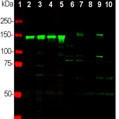
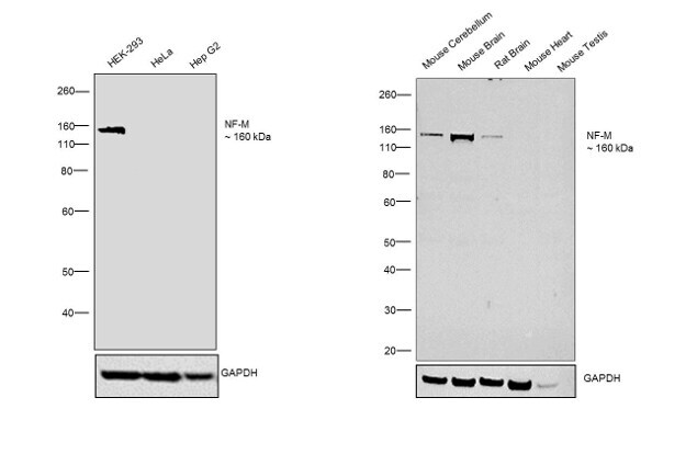
- Western blot was performed using Anti-NEFM Polyclonal Antibody (Product # PA1-16758) and a 160 kDa bands corresponding to NEFM was observed in HEK-293 cell line but not in HeLa and Hep G2. Figure (a) Membrane extracts (30 µg lysate) of HEK-293 (Lane 1), HeLa (Lane 2) and Hep G2 (Lane 3); Figure (b)Tissue extracts (30 µg lysate) of Mouse Cerebellum (Lane 4) Mouse Brain (Lane 5), Rat Brain (Lane 6), Mouse Heart (Lane 7) and Mouse Testis (Lane 8) were electrophoresed using Novex® NuPAGE® 4-12 % Bis-Tris gel (Product # NP0321BOX). Resolved proteins were then transferred onto a nitrocellulose membrane (Product # IB23001) by iBlot® 2 Dry Blotting System (Product # IB21001). The blot was probed with the primary antibody (1:5000 dilution) and detected by chemiluminescence Goat anti-Chicken IgY (H+L) Secondary Antibody, HRP (Product # A16054) using the iBright FL 1000 (Product # A32752). Chemiluminescent detection was performed using Novex® ECL Chemiluminescent Substrate Reagent Kit (Product # WP20005).
Supportive validation
- Submitted by
- Invitrogen Antibodies (provider)
- Main image
- Experimental details
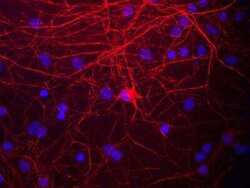
- Immunocytochemistry analysis of NEFM in mixed neuron/glial cultures. Samples were incubated in NEFM polyclonal antibody (Product # PA1-16758). This antibody (red). The NF-M protein is assembled into neurofilaments which are found throughout the axons, dendrites and perikarya of these cells.

- Submitted by
- Invitrogen Antibodies (provider)
- Main image
- Experimental details
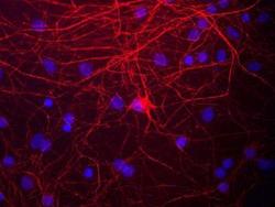
- Immunocytochemistry analysis of NEFM in mixed neuron/glial cultures. Samples were incubated in NEFM polyclonal antibody (Product # PA1-16758). This antibody (red). The NF-M protein is assembled into neurofilaments which are found throughout the axons, dendrites and perikarya of these cells.

- Submitted by
- Invitrogen Antibodies (provider)
- Main image
- Experimental details
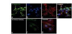
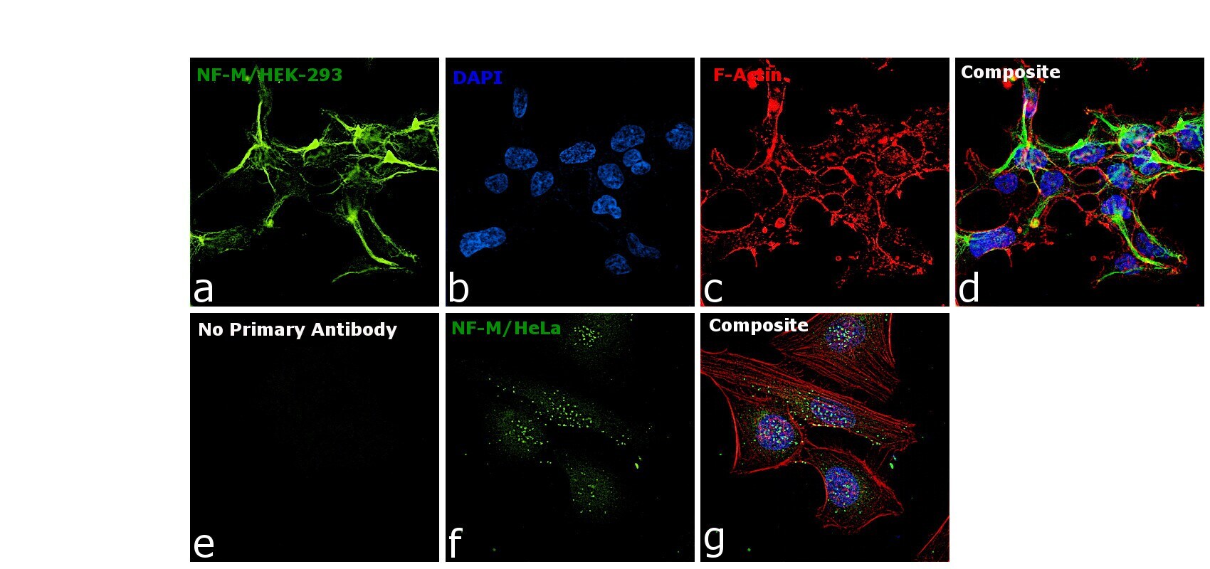
- Immunofluorescence analysis of NF-M was performed using 70% confluent log phase HEK-293 and HeLa cells. The cells were fixed with 4% paraformaldehyde for 10 minutes, permeabilized with 0.1% Triton™ X-100 for 15 minutes, and blocked with 2% BSA for 1 hour at room temperature. HEK-293 cells were labeled with NF-M Chicken Polyclonal Antibody (Product # PA1-16758) at 1:2000 dilution in 0.1% BSA, incubated at 4 degree Celsius overnight and then labeled with Goat anti-Chicken IgY (H+L) Secondary Antibody, Alexa Fluor® 488 conjugate (Product # A-11039) at a dilution of 1:2000 for 45 minutes at room temperature (Panel a: green). Nuclei (Panel b: blue) were stained with ProLong™ Diamond Antifade Mountant with DAPI (Product # P36962). F-actin (Panel c: red) was stained with Rhodamine Phalloidin (Product # R415). Panel d represents the merged image showing cytoskeletal localization. Panel e represents control cells with no primary antibody to assess background. Panel f represents HeLa cells labeled with NF-M Chicken Polyclonal Antibody (Product # PA1-16758) at 1:2000 dilution. Panel g represents the merged image of HeLa cells showing no expression for NF-M protein. The images were captured at 60X magnification.
Supportive validation
- Submitted by
- Invitrogen Antibodies (provider)
- Main image
- Experimental details
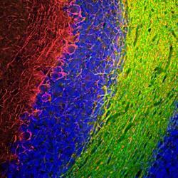
- Immunohistochemical analysis of NEFM in rat cerebellum sections. Samples were incubated in NEFM polyclonal antibody (Product # PA1-16758) using a dilution of 1:1,000 (red). Costained with mouse mAb to CNPase, dilution 1:500, in green. The blue is DAPI staining of nuclear DNA. Following transcardial perfusion of rat with 4% paraformaldehyde, brain was post fixed for 24 hours, cut to 45 µM, and free floating sections were stained with above antibodies. The NF-M antibody labels the axons of basket cells and other neurons, while the CNP antibody stains oligodendrocytes, cells that form the myelin sheathes around axons.
Supportive validation
- Submitted by
- Invitrogen Antibodies (provider)
- Main image
- Experimental details
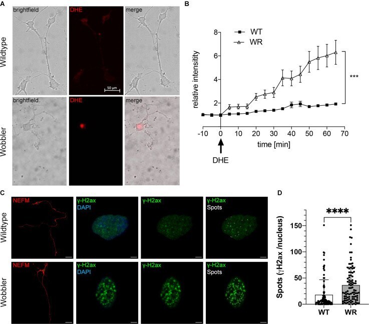
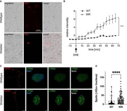
- Elevated ROS levels in motor neurons of Wobbler mice correlate with increased abundance of the DNA damage response protein gammaH2ax. (A) Exemplary images of dissociated motor neurons from wild-type and Wobbler animals (p40+10div) 65 min after DHE (red fluorescence) addition and live cell imaging. Live-cell imaging was performed using a spinning disc confocal microscope with a 20x objective. The fluorescence signals of the cells were documented every 5 min for 1 h in DHE-including medium, 37degC and 5% CO 2 . Scale bar = 50 mum. (B) Relative red fluorescence intensity in five dissociated motor neuronal cell cultures from p40 wild-type and Wobbler mice documented in 5 min intervals of 20 cells per genotype. At timepoint 0 the normal medium was exchanged for DHE including medium. ImageJ 1.53f51 (National Institute of Health, USA) was used to analyze the average red fluorescence intensity within the soma of a motor neuron, while motor neurons themselves were detected in transmitted-light channel. A mean value of background fluorescence intensity without DHE addition was used for normalization. Data are provided as means +- SEM. Data were tested for significance using two-way ANOVA with a Sidak post-hoc test. Significant differences are indicated by *** p < 0.001, n = 20 cells from five dissociated cultures per genotype. (C) Exemplary images of wild-type and Wobbler motor neurons (p40+10div) stained with anti-NEFM antibody (red), anti-phospho-Histone H2A.X (green) and DAPI (blue)