 Explore
 Validate
 Learn
Learn Western blot
Antibody data
- Antibody Data
- Antigen structure
- References [0]
- Comments [0]
- Validations
- Western blot [5]
- Immunocytochemistry [3]
- Immunohistochemistry [1]
Submit
Validation data
Reference
Comment
Report error
- Product number
- PA3-16720 - Provider product page

- Provider
- Invitrogen Antibodies
- Product name
- NEFM Polyclonal Antibody
- Antibody type
- Polyclonal
- Antigen
- Recombinant full-length protein
- Description
- This antibody is likely to react with most mammals.
- Concentration
- Conc. Not Determined
No comments: Submit comment
Supportive validation
- Submitted by
- Invitrogen Antibodies (provider)
- Main image

- Experimental details
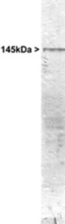
- Western blot of whole rat cerebellum homogenate stained with PA3-16720, at dilution of 1:20,000. A prominent band running with an apparent SDS-PAGE molecular weight of ~145kDa corresponds to rodent NF-M. Human, cow and bovine NF-M run a little slower, at about 160kDa.

- Submitted by
- Invitrogen Antibodies (provider)
- Main image
- Experimental details
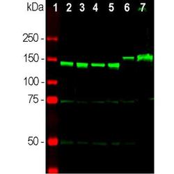
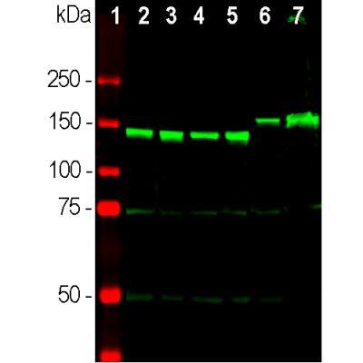
- Western blot analysis of NEFM in neuronal tissue lysates. Samples were incubated in NEFM polyclonal antibody (Product # PA3-16720 using a dilution of 1:2,000. Antibody in green: [1] protein standard (red), [2] rat brain, [3] rat spinal cord, [4] mouse brain, [5] mouse spinal cord, [6] pig brain, [7] pig spinal cord. Strong bands at 145 kDa correspond to rodent NF-M molecules, while the NF-M of pig and other larger mammals including humans run at about 160 kDa.
- Submitted by
- Invitrogen Antibodies (provider)
- Main image
- Experimental details
- Western blot analysis of NEFM in whole rat cerebellum homogenate. Sample was incubated in NEFM polyclonal antibody (Product # PA3-16720).

- Submitted by
- Invitrogen Antibodies (provider)
- Main image

- Experimental details
- Western blot analysis of NEFM was performed by loading 20 µg of HEK-293 wild type (Lane 1), HEK-293 Cas9 control (Lane 2), HEK-293 NEFM knockout (Lane 3) membrane enriched cell extracts. The blot was probed with Anti-NEFM Polyclonal Antibody (Product # PA3-16720) (1:5000 dilution) and Goat anti-Rabbit IgG (H+L), Superclonal™ Recombinant Secondary Antibody, HRP (Product # A27036) (1:4000 dilution). Loss of signal upon CRISPR mediated knockout (KO) confirms that antibody is specific to NEFM.

- Submitted by
- Invitrogen Antibodies (provider)
- Main image
- Experimental details
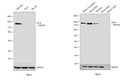
- Western blot was performed using Anti-NEFM Polyclonal Antibody (Product # PA3-16720) and a 160 kDa bands corresponding to NEFM was observed in HEK-293 cell line, Mouse Cerebellum, Mouse Brain and Rat Brain but not in HeLa, Hep G2 cell line, Mouse Heart, Mouse Testis. Figure (a) Membrane extracts (30 µg lysate) of HEK-293 (Lane 1), HeLa (Lane 2) and Hep G2 (Lane 3); Figure (b) Tissue extracts (30 µg lysate) of Mouse Cerebellum (Lane 4), Mouse Brain (Lane 5), Rat Brain (Lane 6), Mouse Heart (Lane 7) and Mouse Testis (Lane 8) were electrophoresed using Novex® NuPAGE® 4-12 % Bis-Tris gel (Product # NP0321BOX). Resolved proteins were then transferred onto a nitrocellulose membrane (Product # IB23001) by iBlot® 2 Dry Blotting System (Product # IB21001). The blot was probed with the primary antibody (1:5000 dilution) and detected by chemiluminescence with Goat anti-Rabbit IgG (H+L), Superclonal™ Recombinant Secondary Antibody, HRP (Product # A27036) Secondary Antibody using the iBright FL 1000 (Product # A32752). Chemiluminescent detection was performed using Novex® ECL Chemiluminescent Substrate Reagent Kit (Product # WP20005).

Supportive validation
- Submitted by
- Invitrogen Antibodies (provider)
- Main image
- Experimental details
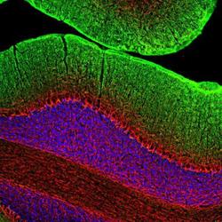
- Immunocytochemistry analysis of NEFM in rat cerebellum section. Samples were incubated in NEFM polyclonal antibody (Product # PA3-16720) using a dilution of 1:2000. This antibody in red, and costained with mouse mAb to GAP43, dilution 1:2,000 in green. Following transcardial perfusion of rat with 4% paraformaldehyde, brain was post fixed for 24 hours, cut to 45 µM, and free-floating sections were stained with the above antibodies. Antibody strongly labels neuronal processes throughout the cerebellum, while the GAP43 antibody stains predominantly synaptic regios in the molecular layer.

- Submitted by
- Invitrogen Antibodies (provider)
- Main image

- Experimental details
- Immunocytochemistry analysis of NEFM in mixed neuron/glia cultures. Samples were incubated in NEFM polyclonal antibody (Product # PA3-16720). NF-M antibody (red) and A2BP1 Antibody (green; mouse monoclonal antibody to Fox1, an mRNA binding protein closely related to Fox3/NeuN). The RPCA-NF-M antibody stains axonal, dendritic and perikaryal profiles of neurons cleanly and specifically. Like antibody to Fox3/NeuN, the Fox1 antibody binds to the nuclei of neurons only. DNA is shown in blue with the DAPI stain.

- Submitted by
- Invitrogen Antibodies (provider)
- Main image
- Experimental details
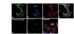
- Immunofluorescence analysis of NF-M was performed using 70% confluent log phase HEK-293 and HeLa cells. The cells were fixed with 4% paraformaldehyde for 10 minutes, permeabilized with 0.1% Triton™ X-100 for 15 minutes, and blocked with 2% BSA for 1 hour at room temperature. HEK-293 cells were labeled with NF-M Rabbit Polyclonal Antibody (Product # PA3-16720) at 1:500 dilution in 0.1% BSA, incubated at 4 degree Celsius overnight and then labeled with Goat anti-Rabbit IgG (H+L), Superclonal™ Recombinant Secondary Antibody, Alexa Fluor 488 (Product # A27034) at a dilution of 1:2000 for 45 minutes at room temperature (Panel a: green). Nuclei (Panel b: blue) were stained with ProLong™ Diamond Antifade Mountant with DAPI (Product # P36962). F-actin (Panel c: red) was stained with Rhodamine Phalloidin (Product # R415). Panel d represents the merged image showing cytoskeletal localization. Panel e represents control cells with no primary antibody to assess background. Panel f represents HeLa cells labeled with NF-M Rabbit Polyclonal Antibody (Product # PA3-16720) at 1:500 dilution. Panel g represents the merged image of HeLa cells showing no expression for NF-M protein. The images were captured at 60X magnification.

Supportive validation
- Submitted by
- Invitrogen Antibodies (provider)
- Main image
- Experimental details
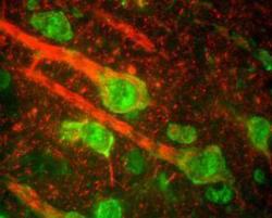
- Immunohistochemical analysis of NEFM in cerebral cortex section from Rat. Frozen samples were incubated in NEFM polyclonal antibody (Product # PA3-16720) using a dilution of 1:5000. This immunostaining reveals the perikarya of pyramidal neurons and dendrites as well as axons surrounding the neurons. Antibody in red and the green channel shows staining with a monoclonal antibody to beta-adrendergic receptor kinase 1.
