 Explore
 Validate
 Learn
Learn Western blot
 Immunocytochemistry
Antibody data
- Antibody Data
- Antigen structure
- References [1]
- Comments [0]
- Validations
- Immunocytochemistry [4]
- Immunohistochemistry [1]
- Other assay [2]
Submit
Validation data
Reference
Comment
Report error
- Product number
- PA5-30442 - Provider product page

- Provider
- Invitrogen Antibodies
- Product name
- U2AF2 Polyclonal Antibody
- Antibody type
- Polyclonal
- Antigen
- Recombinant full-length protein
- Description
- Recommended positive controls: 293T, A431, Jurkat, Raji. Predicted reactivity: Mouse (100%), Xenopus laevis (96%), Pig (100%), Bovine (96%). Store product as a concentrated solution. Centrifuge briefly prior to opening the vial.
- Reactivity
- Human, Mouse
- Host
- Rabbit
- Isotype
- IgG
- Vial size
- 100 μL
- Concentration
- 1.32 mg/mL
- Storage
- Store at 4°C short term. For long term storage, store at -20°C, avoiding freeze/thaw cycles.
Submitted references Alternatively spliced ANLN isoforms synergistically contribute to the progression of head and neck squamous cell carcinoma.
Guo E, Mao X, Wang X, Guo L, An C, Zhang C, Song K, Wang G, Duan C, Zhang X, Yang X, Yuan Z, Sun J, Li X, Yang W, Meng H, Miao S
Cell death & disease 2021 Aug 3;12(8):764
Cell death & disease 2021 Aug 3;12(8):764
No comments: Submit comment
Supportive validation
- Submitted by
- Invitrogen Antibodies (provider)
- Main image
- Experimental details
- U2AF2 Polyclonal Antibody detects U2AF65 protein at nucleus by immunofluorescent analysis. Sample: A431 cells were fixed in 4% paraformaldehyde at RT for 15 min. Green: U2AF65 stained by U2AF2 Polyclonal Antibody (Product # PA5-30442) diluted at 1:500. Red: alpha Tubulin, a cytoskeleton marker, stained by alpha Tubulin Polyclonal Antibody [GT114] (Product # MA5-31466) diluted at 1:1,000.

- Submitted by
- Invitrogen Antibodies (provider)
- Main image

- Experimental details
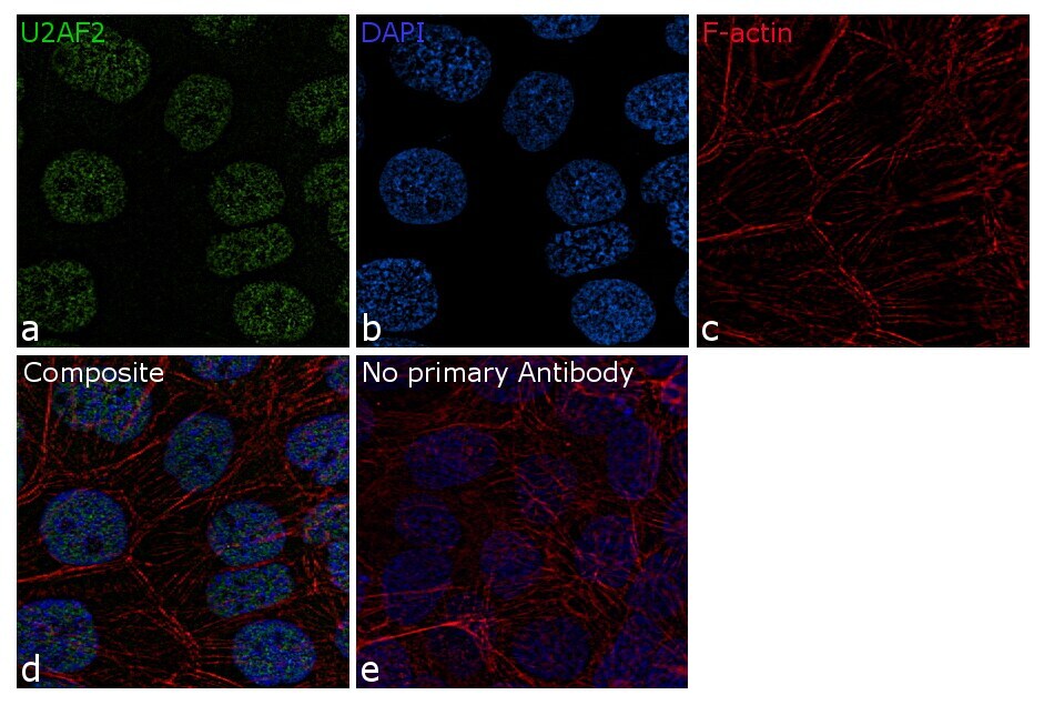
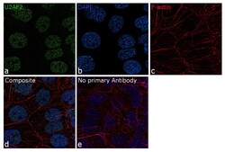
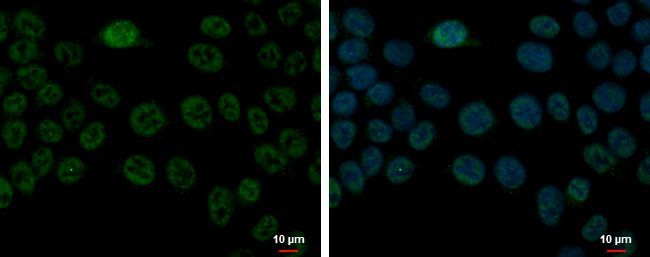
- Immunofluorescence analysis of U2AF2 was performed using 70% confluent A-431 cells. The cells were fixed with 4% paraformaldehyde for 10 minutes, permeabilized with 0.1% Triton™ X-100 for 10 minutes, and blocked with 2% BSA for 1 hour at room temperature. The cells were labeled with U2AF2 Polyclonal Antibody (Product # PA5-30442) at 5µg/mL concentration in 0.1% BSA and incubated overnight at 4 degree and then labeled with Goat anti-Rabbit IgG (H+L) Superclonal™ Secondary Antibody, Alexa Fluor® 488 conjugate (Product # A27034) at a dilution of 1:2000 for 45 minutes at room temperature (Panel a: green). Nuclei (Panel b: blue) were stained with SlowFade® Gold Antifade Mountant with DAPI (Product # S36938). F-actin (Panel c: red) was stained with Rhodamine Phalloidin (Product # R415, 1:300). Panel d and e represents the merged image showing nuclear localization. Panel e represents control cells with no primary antibody to assess background. The images were captured at 60X magnification. .
- Submitted by
- Invitrogen Antibodies (provider)
- Main image
- Experimental details
- Immunofluorescence analysis of U2AF2 was performed using 70% confluent A-431 cells. The cells were fixed with 4% paraformaldehyde for 10 minutes, permeabilized with 0.1% Triton™ X-100 for 10 minutes, and blocked with 2% BSA for 1 hour at room temperature. The cells were labeled with U2AF2 Polyclonal Antibody (Product # PA5-30442) at 5µg/mL concentration in 0.1% BSA and incubated overnight at 4 degree and then labeled with Goat anti-Rabbit IgG (Heavy Chain) Superclonal™ Secondary Antibody, Alexa Fluor® 488 conjugate (Product # A27034) at a dilution of 1:2000 for 45 minutes at room temperature (Panel a: green). Nuclei (Panel b: blue) were stained with SlowFade® Gold Antifade Mountant with DAPI (Product # S36938). F-actin (Panel c: red) was stained with Rhodamine Phalloidin (Product # R415, 1:300). Panel d and e represents the merged image showing nuclear localization. Panel e represents control cells with no primary antibody to assess background. The images were captured at 60X magnification. .

- Submitted by
- Invitrogen Antibodies (provider)
- Main image

- Experimental details
- U2AF2 Polyclonal Antibody detects U2AF65 protein at nucleus by immunofluorescent analysis. Sample: A431 cells were fixed in 4% paraformaldehyde at RT for 15 min. Green: U2AF65 stained by U2AF2 Polyclonal Antibody (Product # PA5-30442) diluted at 1:500. Red: alpha Tubulin, a cytoskeleton marker, stained by alpha Tubulin Polyclonal Antibody [GT114] (Product # MA5-31466) diluted at 1:1,000.

Supportive validation
- Submitted by
- Invitrogen Antibodies (provider)
- Main image
- Experimental details
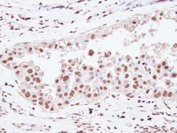
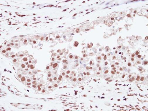
- Immunohistochemical analysis of paraffin-embedded human ovarian cancer, using U2AF65 (Product # PA5-30442) antibody at 1:250 dilution. Antigen Retrieval: EDTA based buffer, pH 8.0, 15 min.
Supportive validation
- Submitted by
- Invitrogen Antibodies (provider)
- Main image
- Experimental details
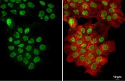
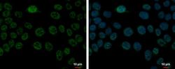
- Immunofluorescent analysis of U2AF65 showing staining in the nucleus of HCT 116 cells. HCT 116 cells were fixed in 4% paraformaldehyde at RT for 15 min and stained using a U2AF65 polyclonal antibody (Product # PA5-30442) diluted at 1:500. Blue: Hoechst 33342 staining. Scale bar = 10µm.
- Submitted by
- Invitrogen Antibodies (provider)
- Main image
- Experimental details
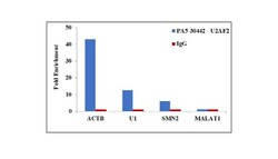
- RNA Immunoprecipitation (RIP) assay of endogenous U2AF2 protein using Anti-U2AF2 Antibody: RIP assay was performed using Anti-U2AF2 Recombinant Rabbit Polyclonal Antibody (Product # PA5-30442, 5 ug) on whole cell lysate from Hep G2 cells. Normal Rabbit IgG was used as a negative IP control. RNA purified by RiboPure™ RNA Purification Kit (Product # AM1924) was analyzed by RT-PCR using the Power SYBR® Green RNA-to-CT™ 1-Step Kit (Product # 4389986) with the primers pairs over ACTB, SMN2 mRNA and MALAT1 non-coding RNA. Data is presented as fold enrichment of the antibody signal versus the negative control IgG using the comparative CT method.
