Explore
 Validate
 Learn
Learn Western blot
Antibody data
- Antibody Data
- Antigen structure
- References [1]
- Comments [0]
- Validations
- Western blot [1]
- Immunohistochemistry [1]
Submit
Validation data
Reference
Comment
Report error
- Product number
- MAB7447 - Provider product page

- Provider
- R&D Systems
- Product name
- Human Urocortin Antibody
- Antibody type
- Monoclonal
- Description
- Protein A or G purified from hybridoma culture supernatant. Detects human Urocortin in direct ELISAs and Western blots.
- Reactivity
- Human
- Host
- Mouse
- Conjugate
- Unconjugated
- Antigen sequence
P55089- Isotype
- IgG
- Antibody clone number
- 749606
- Vial size
- 100 ug
- Concentration
- LYOPH
- Storage
- Use a manual defrost freezer and avoid repeated freeze-thaw cycles. 12 months from date of receipt, -20 to -70 °C as supplied. 1 month, 2 to 8 °C under sterile conditions after reconstitution. 6 months, -20 to -70 °C under sterile conditions after reconstitution.
Submitted references Urocortin suppresses endometrial cancer cell migration via CRFR2 and its system components are differentially modulated by estrogen.
Owens GL, Lawrence KM, Jackson TR, Crosbie EJ, Sayan BS, Kitchener HC, Townsend PA
Cancer medicine 2017 Feb;6(2):408-415
Cancer medicine 2017 Feb;6(2):408-415
No comments: Submit comment
Supportive validation
- Submitted by
- R&D Systems (provider)
- Main image
- Experimental details
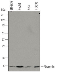
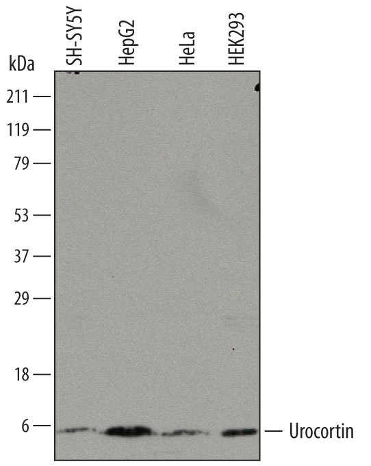
- Detection of Human Urocortin by Western Blot. Western blot shows lysates of SH-SY5Y human neuroblastoma cell line, HepG2 human hepatocellular carcinoma cell line, HeLa human cervical epithelial carcinoma cell line, and HEK293 human embryonic kidney cell line. PVDF membrane was probed with 0.5 µg/mL of Mouse Anti-Human Urocortin Monoclonal Antibody (Catalog # MAB7447) followed by HRP-conjugated Anti-Mouse IgG Secondary Antibody (Catalog # HAF007). A specific band was detected for Urocortin at approximately 5 kDa (as indicated). This experiment was conducted under reducing conditions and using Immunoblot Buffer Group 8.
Supportive validation
- Submitted by
- R&D Systems (provider)
- Main image

- Experimental details
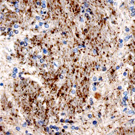
- Urocortin in Human Brain. Urocortin was detected in immersion fixed paraffin-embedded sections of human brain using Mouse Anti-Human Urocortin Monoclonal Antibody (Catalog # MAB7447) at 15 µg/mL overnight at 4 °C. Before incubation with the primary antibody, tissue was subjected to heat-induced epitope retrieval using Antigen Retrieval Reagent-Basic (Catalog # CTS013). Tissue was stained using the Anti-Mouse HRP-DAB Cell & Tissue Staining Kit (brown; Catalog # CTS002) and counterstained with hematoxylin (blue). Specific staining was localized to neuronal processes. View our protocol for Chromogenic IHC Staining of Paraffin-embedded Tissue Sections.