 Explore
 Validate
 Learn
Learn Western blot
 Immunocytochemistry
Antibody data
- Antibody Data
- Antigen structure
- References [1]
- Comments [0]
- Validations
- Immunocytochemistry [3]
- Other assay [1]
Submit
Validation data
Reference
Comment
Report error
- Product number
- PA5-77985 - Provider product page

- Provider
- Invitrogen Antibodies
- Product name
- GFI1 Polyclonal Antibody
- Antibody type
- Polyclonal
- Antigen
- Recombinant full-length protein
- Description
- Positive Control: THP-1, HL-60 Predicted Reactivity: Mouse (80%), Rat (81%), Dog (87%), Cat (89%), Rhesus Monkey (98%), Bovine (88%) Store product as a concentrated solution. Centrifuge briefly prior to opening the vial.
- Reactivity
- Human
- Host
- Rabbit
- Isotype
- IgG
- Vial size
- 100 μL
- Concentration
- 1.49 mg/mL
- Storage
- Store at 4°C short term. For long term storage, store at -20°C, avoiding freeze/thaw cycles.
Submitted references An optimized permeabilization step for flow cytometry analysis of nuclear proteins in myeloid differentiation of blood cells into neutrophils.
Viryasova GM, Golenkina EA, Tatarskii VV Jr, Galkin II, Sud'ina GF, Soshnikova NV
MethodsX 2019;6:360-367
MethodsX 2019;6:360-367
No comments: Submit comment
Supportive validation
- Submitted by
- Invitrogen Antibodies (provider)
- Main image
- Experimental details
- Immunocytochemistry-Immunofluorescence analysis of GFI1 was performed in THP-1 cells fixed in 4% paraformaldehyde at RT for 15 min. Green: GFI1 Polyclonal Antibody (Product # PA5-77985) diluted at 1:500. Red: phalloidin, a cytoskeleton marker. Scale bar = 10 µm.

- Submitted by
- Invitrogen Antibodies (provider)
- Main image

- Experimental details
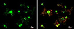
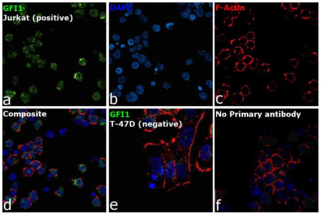
- Immunofluorescence analysis of GFI-1 was performed in Jurkat and T-47D cells. The cells were fixed with 4% paraformaldehyde for 10 minutes, permeabilized with 0.1% Triton™ X-100 for 15 minutes, and blocked with 2% BSA for 1 hour at room temperature. The cells were labeled with GFI1 Polyclonal Antibody (Product # PA5-77985) at 1:100 dilution in 0.1% BSA, incubated at 4 degree celsius overnight and then labeled with Donkey anti-Rabbit IgG (H+L) Highly Cross-Adsorbed Secondary Antibody, Alexa Fluor™ Plus 488 (Product # A32790, 1:2000 dilution), for 45 minutes at room temperature (Panel a: Green). Nuclei (Panel b:Blue) were stained with ProLong™ Diamond Antifade Mountant with DAPI (Product # P36962). F-actin (Panel c: Red) was stained with Rhodamine Phalloidin (Product # R415, 1:300). Panel d represents the merged image showing Nuclear localization. Panel e represents no signal in the negative model (T-47D). Panel f represents control cells with no primary antibody to assess background. The images were captured at 60X magnification.
- Submitted by
- Invitrogen Antibodies (provider)
- Main image

- Experimental details
- Immunofluorescence analysis of GFI-1 was performed in Jurkat and T-47D cells. The cells were fixed with 4% paraformaldehyde for 10 minutes, permeabilized with 0.1% Triton™ X-100 for 15 minutes, and blocked with 2% BSA for 1 hour at room temperature. The cells were labeled with GFI1 Polyclonal Antibody (Product # PA5-77985) at 1:100 dilution in 0.1% BSA, incubated at 4 degree celsius overnight and then labeled with Donkey anti-Rabbit IgG (H+L) Highly Cross-Adsorbed Secondary Antibody, Alexa Fluor™ Plus 488 (Product # A32790, 1:2000 dilution), for 45 minutes at room temperature (Panel a: Green). Nuclei (Panel b:Blue) were stained with ProLong™ Diamond Antifade Mountant with DAPI (Product # P36962). F-actin (Panel c: Red) was stained with Rhodamine Phalloidin (Product # R415, 1:300). Panel d represents the merged image showing Nuclear localization. Panel e represents no signal in the negative model (T-47D). Panel f represents control cells with no primary antibody to assess background. The images were captured at 60X magnification.

Supportive validation
- Submitted by
- Invitrogen Antibodies (provider)
- Main image
- Experimental details
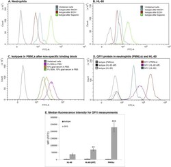
- Fig. 2 Flow cytometry overlay diagrams demonstrate AlexaFluor488-labelled antibodies distribution in neutrophils after permeabilization with 70% EtOH, 90% MeOH or 0.1% saponin. (A) The efficiency of block of secondary Ab non-specific binding (fluorescence level of isotype control) depending on the permeabilization agents in human leukocytes, primary cells. (B) Isotype controls in case of different permeabilization agents in HL-60 cells. (C) Isotype controls in PMNLs after possible non-specific binding sites were blocked with different reagents: 1% BSA, 10% goat serum or both. (D) The alterations of GFI1 protein content during myeloid differentiation of cell line HL-60 with trans-retinoic acid (ATRA) and in human neutrophils, samples were prepared according to the Protocol. (E) The median fluorescence intensity data for GFI1 protein. Values indicate the mean +- SD from three independent experiments. ** p < 0.01, *** p < 0.005 compared to the GFI1 content in HL-60. For all pairs Isotype-GFI1b p-value is less than 0.005 (not mentioned on the diagram. Statistical analysis was done with 2-way ANOVA, Holm-Sidak's multiple comparison test. Fig. 2
