Explore
 Validate
 Learn
Learn Western blot
 Immunocytochemistry
Antibody data
- Antibody Data
- Antigen structure
- References [2]
- Comments [0]
- Validations
- Immunocytochemistry [1]
Submit
Validation data
Reference
Comment
Report error
- Product number
- HPA028425 - Provider product page

- Provider
- Atlas Antibodies
- Proper citation
- Atlas Antibodies Cat#HPA028425, RRID:AB_10600899
- Product name
- Anti-CSK
- Antibody type
- Polyclonal
- Description
- Polyclonal Antibody against Human CSK, Gene description: c-src tyrosine kinase, Validated applications: WB, IHC, ICC, Uniprot ID: P41240, Storage: Store at +4°C for short term storage. Long time storage is recommended at -20°C.
- Reactivity
- Human, Mouse, Rat
- Host
- Rabbit
- Conjugate
- Unconjugated
- Isotype
- IgG
- Vial size
- 100 µl
- Concentration
- 0.2 mg/ml
- Storage
- Store at +4°C for short term storage. Long time storage is recommended at -20°C.
- Handling
- The antibody solution should be gently mixed before use.
Submitted references GDF6-CD99 Signaling Regulates Src and Ewing Sarcoma Growth.
Beta-catenin inhibits melanocyte migration but induces melanoma metastasis
Zhou F, Elzi DJ, Jayabal P, Ma X, Chiu YC, Chen Y, Blackman B, Weintraub ST, Houghton PJ, Shiio Y
Cell reports 2020 Nov 3;33(5):108332
Cell reports 2020 Nov 3;33(5):108332
Beta-catenin inhibits melanocyte migration but induces melanoma metastasis
Gallagher S, Rambow F, Kumasaka M, Champeval D, Bellacosa A, Delmas V, Larue L
Oncogene 2012;32(17):2230-2238
Oncogene 2012;32(17):2230-2238
No comments: Submit comment
Supportive validation
- Submitted by
- Atlas Antibodies (provider)
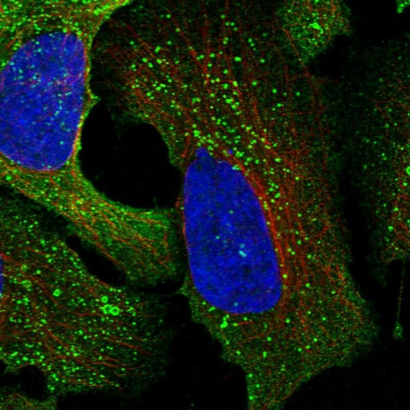
- Main image

- Experimental details
- Immunofluorescent staining of human cell line U-2 OS shows localization to cytosol & vesicles.
- Sample type
- Human