Explore
 Validate
 Learn
LearnOAAB03940
antibody from Aviva Systems Biology
Targeting: PTP4A2
HU-PP-1, OV-1, PRL-2, PRL2, ptp-IV1a, PTP4A, PTPCAAX2
 Western blot
 ELISA
ELISAAntibody data
- Antibody Data
- Antigen structure
- References [0]
- Comments [0]
- Validations
- Western blot [1]
- Immunohistochemistry [2]
- Flow cytometry [2]
Submit
Validation data
Reference
Comment
Report error
- Product number
- OAAB03940 - Provider product page

- Provider
- Aviva Systems Biology
- Product name
- PTP4A2 antibody - center region
- Antibody type
- Polyclonal
- Reactivity
- Human
- Host
- Rabbit
- Vial size
- 400ul
- Storage
- Maintain refrigerated at 2-8 deg C for up to 6 months. For long term storage store at -20 deg C in small aliquots to prevent freeze-thaw cycles.
No comments: Submit comment
Supportive validation
- Submitted by
- Aviva Systems Biology (provider)
- Main image

- Experimental details
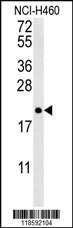
- PTP4A2 Antibody (Center) western blot analysis in NCI-H460 cell lysates (35ug/lane).This demonstrates the PTP4A2 antibody detected the PTP4A2 protein (arrow).
- Sample type
- NCI-H460 cell line lysates
- Primary Ab dilution
- 1.0 µg/mL
- Protocol
- Protocol
Supportive validation
- Submitted by
- Aviva Systems Biology (provider)
- Main image

- Experimental details
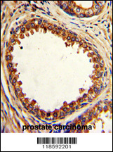
- Formalin-fixed and paraffin-embedded human prostate carcinoma with PTP4A2 Antibody (Center), which was peroxidase-conjugated to the secondary antibody, followed by DAB staining. This data demonstrates the use of this antibody for immunohistochemistry; clinical relevance has not been evaluated.
- Sample type
- human prostate carcinoma tissue
- Primary Ab dilution
- 1.0 µg/mL
- Protocol
- Protocol
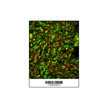
- Submitted by
- Aviva Systems Biology (provider)
- Main image
- Experimental details
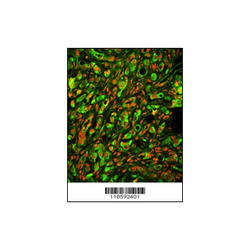
- Immunofluorescence analysis of PTP4A2 Antibody (Center) with paraffin-embedded human prostate carcinoma tissue . 0.05 mg/ml primary antibody was followed by FITC-conjugated goat anti-rabbit lgG (whole molecule). FITC emits green fluorescence.Red counterst
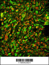
Supportive validation
- Submitted by
- Aviva Systems Biology (provider)
- Main image
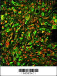
- Experimental details
- Immunofluorescence analysis of PTP4A2 Antibody (Center) with paraffin-embedded human prostate carcinoma tissue . 0.05 mg/ml primary antibody was followed by FITC-conjugated goat anti-rabbit lgG (whole molecule). FITC emits green fluorescence.Red counterst
- Sample type
- human prostate carcinoma tissue
- Primary Ab dilution
- 1.0 µg/mL
- Protocol
- Protocol
- Submitted by
- Aviva Systems Biology (provider)
- Main image

- Experimental details
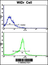
- Flow cytometric analysis of widr cells using PTP4A2 Antibody (Center)(bottom histogram) compared to a negative control cell (top histogram). FITC-conjugated goat-anti-rabbit secondary antibodies were used for the analysis.
- Sample type
- widr cells
- Primary Ab dilution
- 1.0 µg/mL
- Protocol
- Protocol